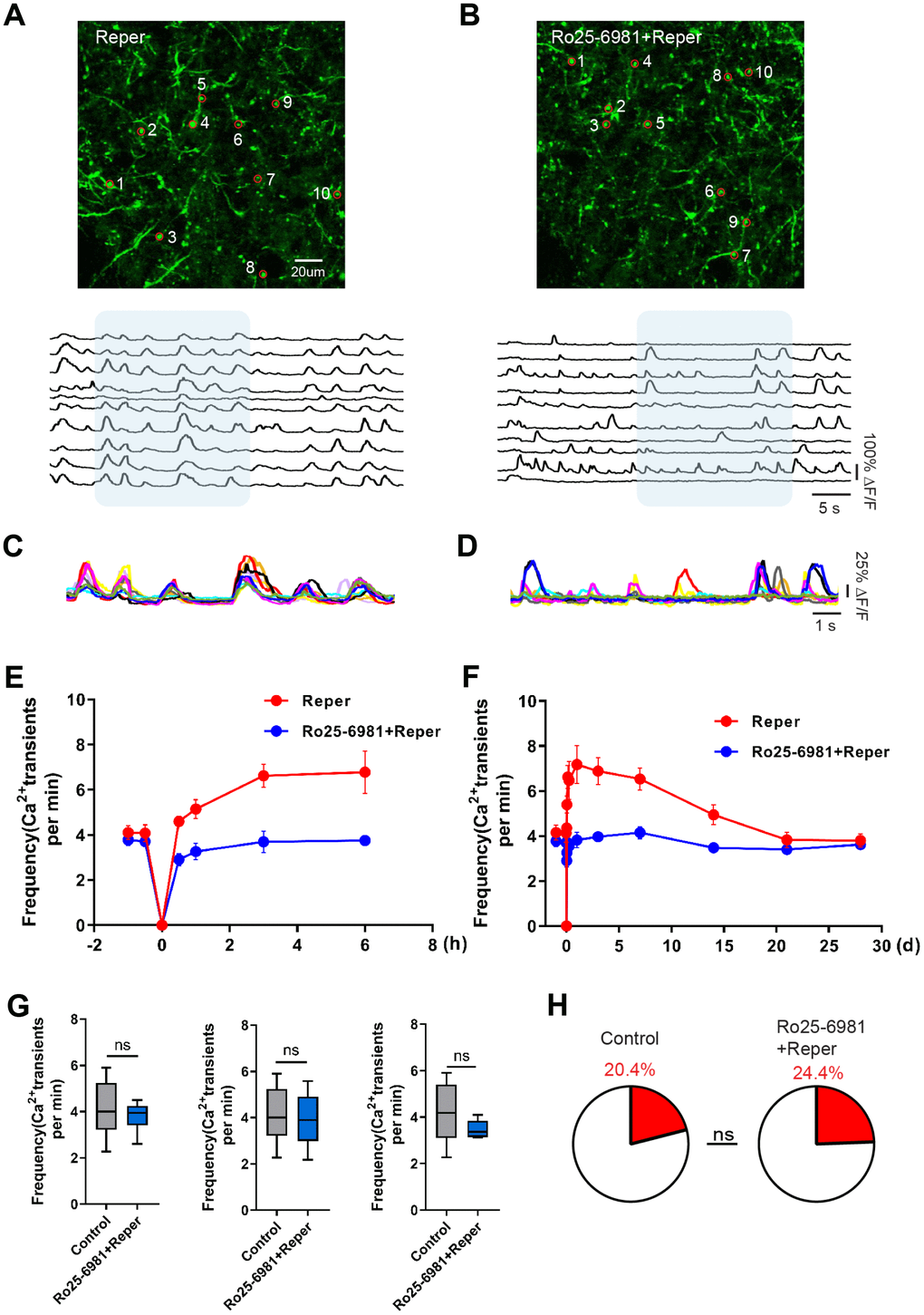
Figure 2.Transient global cerebral ischemia-reperfusion aggravates NMDAR-dependent spine dysfunction. (A, B) Top, layer 1 (30~50 um) spines images on the 3rd day after reperfusion, where the red circle represents the selected spine in Reper (A) and Ro25-6981-treated (B) mice. Bottom, the traces of selected spines. (C, D) Superimposed traces from the shaded areas in a and b. Each color represents a different cell. (E, F) The overall trend of the average frequency of Ca2+ transients before and after 6 hours (E) and 28 days (F) of ischemia-reperfusion. (G) The average frequency of Ca2+ transients in Reper and Ro25-698-treated mice. The 3rd hour (left, n= 438 spines in 6 Reper mice, n= 312 spines in 4 Ro25-698-treated mice), the 3rd day (middle, n= 432 spines in 6 Reper mice, n= 328 spines in 4 Ro25-698-treated mice) and the 14th day (right, n=436 spines in 6 Reper mice, n= 329 spines in 4 Ro25-6981-treated mice). (H) The fractions of hyperactive spines Reper mice (n= 432 spines) and Ro25-698-treated mice (n=328 spines) on the 3rd day after reperfusion. *P < 0.05, ns, not significant, Student’s t-test. Error bars = s.e.m.