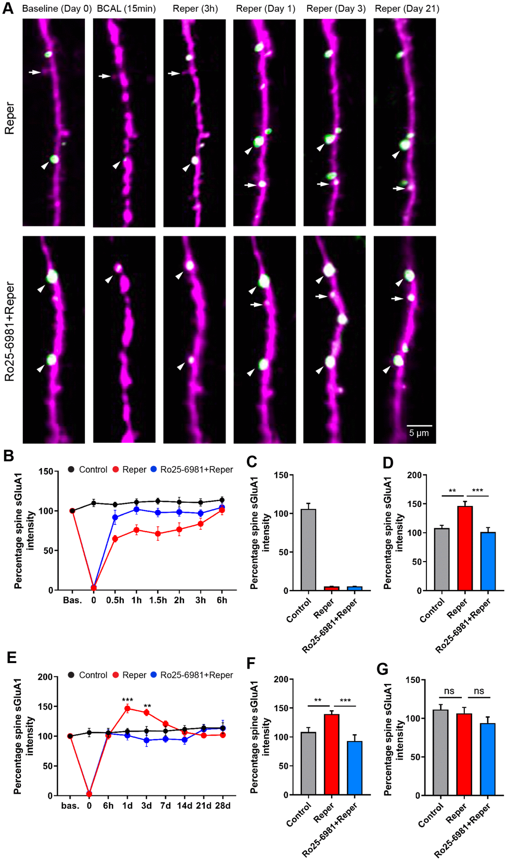
Figure 5.BCAL leads to an NMDAR-dependent increase in spine sGluA1 in vivo in apical dendrites of layer 2/3 neurons in the somatosensory cortex. (A) Representative images of spines on layer 2/3 apical dendrites in Reper or Ro25-6981 treated mice. Arrowheads indicate stable spines and arrows mark unstable spines, including new spines and eliminated spines. (B, E) The overall trend of the Spine sGluA1 intensity in control, Reper and Ro25-6981-treated mice before and after 6 hours (B) and 28 days (E) of ischemia-reperfusion. **P < 0.01, ***P < 0.001, two-way ANOVA with Bonferroni correction. Error bars= s.e.m. (C–G) Percentage spine sGluA1 at the beginning of reperfusion (C), on the first day (D, n= 425 spines in 5 control mice, n= 379 spines in Reper mice, and n= 411 spines in 5 Ro25-6981-treated mice.), the 3rd day (F, n= 410 spines in 5 control mice, n= 283 spines in Reper mice, and n= 274 spines in 5 Ro25-6981-treated mice.) and the 14th day (G, n= 410 spines in 5 control mice, n= 283 spines in Reper mice, and n= 274 spines in 5 Ro25-6981-treated mice.) following ischemia-reperfusion. **P < 0.01, ***P < 0.001, ns, not significant, one-way ANOVA with Bonferroni correction. Error bars = s.e.m.