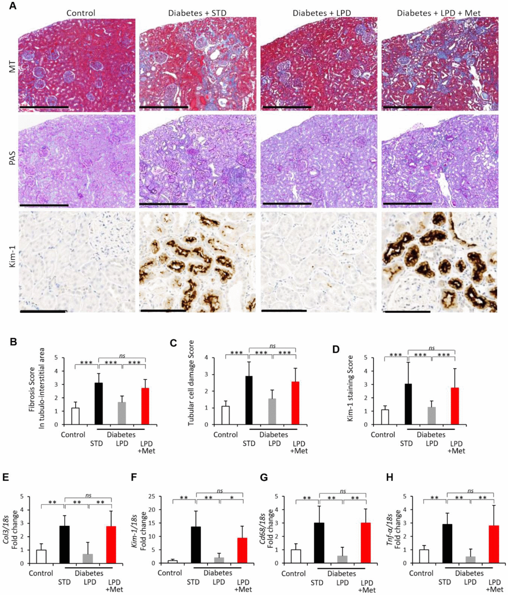
Figure 3.Changes in renal fibrosis and tubular cell damage. Representative photographs of MT, PAS staining and Kim-1 immunohistochemistry of the kidney after the intervention. MT staining of the tubulointerstitial area (scale bar: 500 μm), PAS staining for the evaluation of tubular cell damage (scale bar: 500 μm) and immunohistochemistry for Kim-1 (scale bar: 100 μm) at the end of the study (A); Tubulointerstitial fibrotic scores obtained using MT staining (B) (n=7); tubular cell damage scores obtained using PAS staining (C) (n=7); semiquantitation of Kim-1 staining scores (D) (n=3); mRNA expression of Col3 (E), Kim-1 (F), Cd68 (G) and Tnf-α (H) adjusted to 18S levels, in the renal cortex at the end of the study (n=7). The data shown are the means ± SD. *p<0.05, **p<0.01, ***p<0.001 vs. the indicated groups. ns: not significant. MT: Masson’s trichrome, PAS: Periodic Acid Schiff, Col3: type 3 collagen, Kim-1: kidney injury molecule-1, Tnf-α: tumor necrosis factor-α.