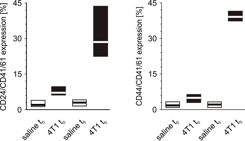
Figure 11.The formation of platelet-cancer cells aggregates in mice injected with 4T1 cells or saline. Results are presented as median (horizontal line) and interquartile range (box) (n = 8). The expressions of CD24 or CD44 within the population of the CD41/61-gated cells (platelets) were measured using flow cytometry in non-fixed ‘washed blood’ drawn immediately (t0) or after 5 weeks (t5) from the injection of 4T1 cells (n = 8). Results are expressed as the percent fraction of CD24/CD41/61 or CD44/CD41/61-positive cells. For further experimental details – see Materials and methods section. The statistical significance of differences, estimated with Kruskal-Wallis test followed by post hoc Conover-Inman all-pairwise comparisons or one-way ANOVA followed by Tukey’s multiple comparisons test, was: CD24/CD41/61-positive cells (4T1-platelet aggregates): P1,α < 0.001, 4T1 t0 > saline t0, 4T1 t5 > saline t5; CD44/CD41/61-positive cells (4T1-platelet aggregates): P1,α < 0.01, 4T1 t0 > saline t0; P1,α < 0.01, 4T1 t5 > saline t5.