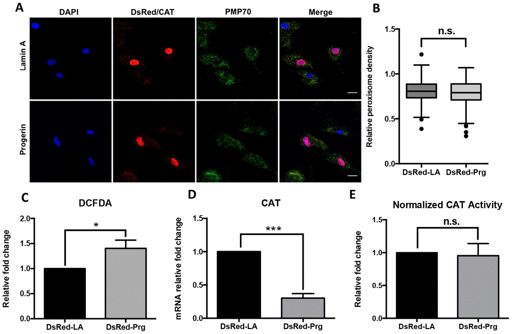
Figure 3.Peroxisomal defects in normal fibroblasts expressing progerin. (A) Normal human dermal fibroblast cells were infected by DsRed-LA and DsRed-Pg lentiviruses. The exogenous Lamin A and progerin expression were detected by DsRed fluorescence in the nuclei. Peroxisomes localization was indicated by PMP70 and catalase immunofluorescence staining. Bar = 25μm. (B) Quantification of PMP70 puncta per square unit in fibroblasts overexpressing lamin A and progerin. More than 200 cells from 3 independent experiments were analyzed and the data was represented in Tukey box plot. Boxes show the 25th, 50th, and 75th percentiles and the dots indicate the outliers. (C) Relative fold change of ROS activity measured by DCFDA flow cytometry analysis in fibroblasts overexpressing lamin A and progerin. *, p < 0.05. (D) Quantitative RT-PCR analysis of the relative expression of catalase in fibroblasts overexpressing lamin A and progerin. ***, p < 0.001. (E) Normalized catalase activity in fibroblasts overexpressing lamin A and progerin. n.s., not significant. All experiments were performed using mid-passage cells between p15 to p25. All experiments were repeated at least three times and representative data were shown as indicated.