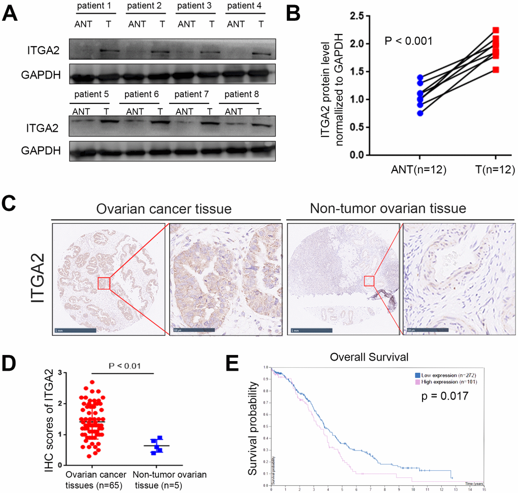
Figure 1.The overexpressed ITGA2 is correlated with poor prognosis in ovarian cancer. (A, B) Western blot assay was conducted to explore the protein expression of ITGA2 in 8 paired primary ovarian cancer tissues (T) and the matched adjacent normal tissues (ANT) of the same patient (A). The quantified proteins expression level of ITGA2 were also shown (B). The P values were shown in the figure. Statistical analyses were performed with D’Agostino and Pearson omnibus normality test. (C) TMA tissue sections were used for ITGA2 IHC staining. The IHC images were shown. The scale bars were shown in the figure. (D) Dot plots to show the IHC score of ITGA2 expression using TMA tissue sections (normal ovarian specimens: n = 5, ovarian cancer TMA specimens: n = 65, P < 0.001). Statistical analyses were performed with D’Agostino and Pearson omnibus normality test. (E) The overall survival of ovarian cancer patients was searched by Human Protein Atlas database (P < 0.001).