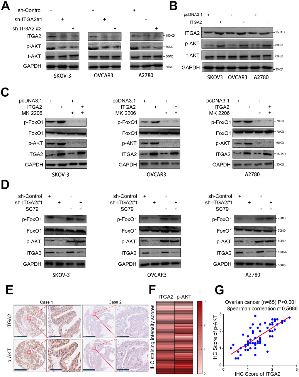
Figure 4.ITGA2 contributes to activating AKT pathway in ovarian cancer cells. (A) SKOV3, OVCAR3, and A2780 cells lines were infected with sh-Control, sh-ITGA2 #1, or sh-ITGA2 #2. Western blot analysis was performed with these cells after 72 hours culturing. GAPDH served as an internal reference. (B) SKOV3, OVCAR3, and A2780 cells lines were infected with or without ITGA2 plasmids. Western blot analysis was performed with these cells after 72 hours culturing. GAPDH served as an internal reference. (C) SKOV3, OVCAR3, and A2780 cells lines were infected with or without ITGA2 plasmids and were treated with or without AKT inhibitor (MK 2206). Western blot analysis was performed with these cells after 72 hours culturing. GAPDH served as an internal reference. (D) SKOV3, OVCAR3, and A2780 cells lines were infected with sh-Control, sh-ITGA2 #1, or sh-ITGA2 #2 and were treated with or without AKT agonist (SC79). Western blot analysis was performed with these cells after 72 hours culturing. GAPDH served as an internal reference. (E) IHC Images of ITGA2 and p-AKT staining using TMA tissue sections (n = 65 ovarian cancer patient specimens). The scale bars were shown as indicated. f and g. Heatmap (F) and dot plot (G) to show the correlation of IHC scores for the expression of the ITGA2 and p-AKT proteins in ovarian cancer patient specimens. (r = 0.5686 for spearman correlation coefficients, P < 0.001).