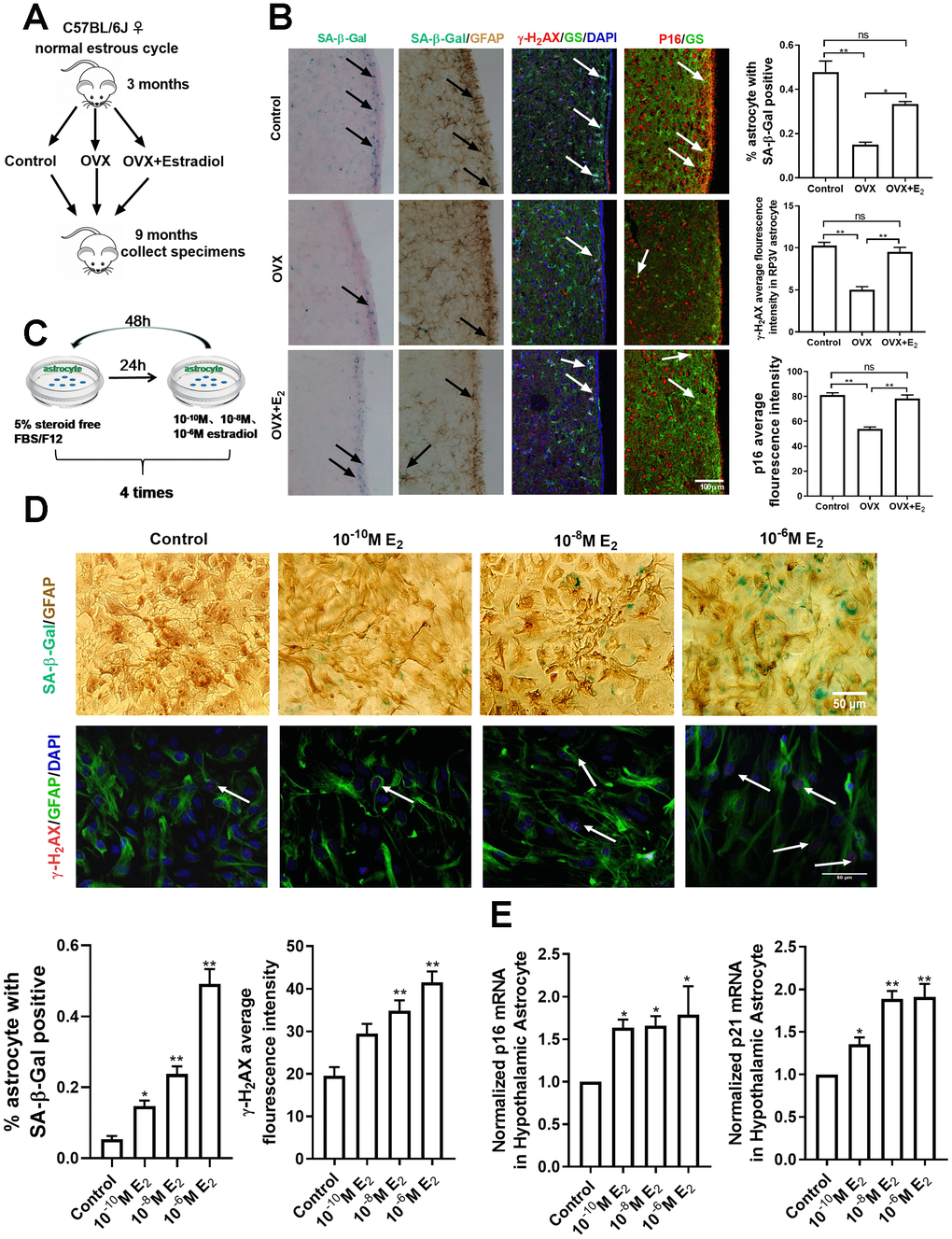
Figure 2.Estradiol induces senescence of astrocytes in the hypothalamus. (A) The flow chart of mouse castration and estradiol intervention. (B) Representative microscopies showing SA-β-gal staining in the control (n=5), OVX (n=5) and OVX+E2 groups (n=5), black arrows representing SA-β-Gal–positive cells (left one). Dual-label immunohistochemistry showing astrocytes by GFAP staining (brown) and by SA-β-Gal staining (blue), black arrows representing SA-β-Gal–positive astrocytes (left two). Dual-label immunofluorescence showing astrocytes (green) with γ-H2AX (red), white arrows representing γ-H2AX–positive astrocytes (right two). Dual-label immunofluorescence showing astrocytes (green) with p16 (red), white arrows representing p16–positive astrocytes (right one). Scale bar= 100μm. (C) The flow chart of estradiol intervention in primary cultured astrocytes. (D) Dual-label immunohistochemistry showing astrocytes by GFAP staining (brown) and by SA-β-Gal staining (blue) with three different estradiol concentrations (10-10M, 10-8M, 10-6M) (upper), black arrows representing SA-β-Gal–positive astrocytes. Scale bar= 100 μm. Dual-label immunofluorescence showing astrocytes (green) and γ-H2AX (red) with different estradiol concentrations, white arrows representing γ-H2AX–positive astrocytes. Scale bar=50μm. (E) Detection of p16 and p21 mRNA levels under different estradiol concentrations. Estradiol increased the expression of p16 and p21 with different estradiol concentrations in hypothalamic astrocytes (n = 3-4). The experiments used two-way analysis of variance. The p-value was determined by One-way ANOVA: *p<0.05, ** p< 0.01.OVX, i.e. ovariectomy, OVX+E2, i.e. ovariectomy plus estradiol replacement, 10-6M E2, i.e.10-6M estradiol concentrations.