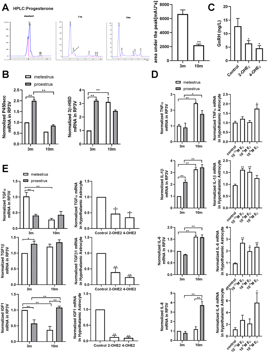
Figure 5.Estradiol-induced senescence of hypothalamic astrocytes contributes to aging-related declines in female reproductive function. (A) HPLC chromatogram of standard progesterone (left) and hypothalamic tissue extract of 3-month-old and 10-month-old mice. The p-value was determined by Student’s t test: ** p<0.01, n=3. (B) Effects of estrous cycle and age on the levels of P450scc and 3β-HSD mRNA in hypothalamus as determined by qPCR. The p-value was determined by Two-way ANOVA: ** p< 0.01, n = 3. (C) Both 2-OHE2-ACM and 4-OHE2-ACM inhibited the secretion of GnRH from GT1-7 cells. The p-value was determined by One-way ANOVA: *p<0.05, n=3-4. (D) Effects of estrous cycle and age on the levels of TNF-α, IL-1β, IL-6 and IL-8 mRNA in hypothalamus as determined by qPCR. The p-value was determined by Two-way ANOVA: *p<0.05, ** p< 0.01, n = 3 (upper). Effects of estradiol on the levels of TNF-α, IL-1β, IL-6 and IL-8 mRNA in hypothalamic primary cultured astrocytes as determined by qPCR (n=3-6). The p-value was determined by One-way ANOVA:*p<0.05, ** p< 0.01 (blew). (E) Effects of estrous cycle and age on the levels of TGF-α, TGF-β1, and IGF1 mRNA in hypothalamus as determined by qPCR (n = 3). The p-value was determined by Two-way ANOVA: *p<0.05, ** p< 0.01 (upper). Detection of TGF-α, TGF-β1, and IGF1 mRNA levels with 2-OHE2 and 4-OHE2 intervention (n=3). The p-value was determined by One-way ANOVA:*p<0.05, ** p< 0.01.