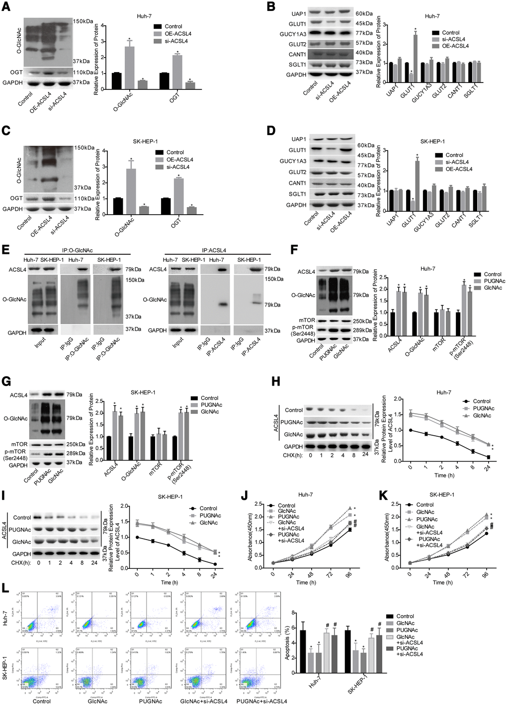
Figure 3.Evaluation of the effects of ACSL4 on O-GlcNAc-mediated HCC growth. Huh-7 and SK-HEP-1 cells were transfected with OE-ACSL4 or si-ACSL4, and then the cells were harvested for the western blotting assay to detect the expression of the following proteins. (A) OGT and O-GlcNAc in Huh-7 cells. (B) UAP1, GLUT1, GLUT2, GUCY1A3, CANT1 and SGLT1 in Huh-7 cells. (C, D) OGT, O-GlcNAc, UAP1, GLUT1, GLUT2, GUCY1A3, CANT1 and SGLT1 in SK-HEP-1 cells. (E) IP assay used to detect the interaction between O-GlcNAc and ACSL4 with an antibody against O-GlcNAc or ACSL4. IgG served as a negative control. Then, the si-ACSL4-transfected or untransfected Huh-7 and SK-HEP-1 cells were treated with PUGNAc, GlcNAc or nothing, and the following assays were carried out. (F, G) The levels of ACSL4, O-GlcNAc, mTOR and p-mTOR were determined by using a western blotting assay. (H, I) The protein stability was determined by western blotting after incubation with CHX (100 μg/ml) for 0, 1, 2, 4, 8 or 24 hours. (J, K) Cell proliferation was detected by CCK-8 assay. (L) Cell apoptosis was assessed by flow cytometry assay. (A–D) *P<0.05, si-ACSL4/OE-ACSL4 group compared with control group; (E–L)*P<0.05, PUGNAc/GlcNAc group compared with control group; #P<0.05, PUGNAc + si-ACSL4/GlcNAc + si-ACSL4 group compared with PUGNAc/GlcNAc group).