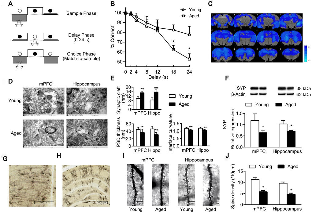
Figure 1.Cognition changes in aged rats. (A) The DMTP procedure, consisting of sample phase, delay phase and choice phase. ○ illumination of the stimulus light or panel light, ● extinguished stimulus light or panel light. (B) Cognitive performance of young and aged rats analyzed by DMTP; n = 12. (C) Images of brain slices showing regions with lower ReHo in aged rats compared with young rats; n = 20. (voxel level < 0.005, cluster level < 0.05 GRF corrected, and clusters > 50 voxels). Blue denotes lower ReHo; the color bars indicated the T values between groups. (D) The synaptic structures of mPFC and hippocampus in young and aged rats by transmission electron microscopy (× 60000). n = 3. (E) Histograms of synaptic structure parameters. n = 3. (F) Expression of synaptophysin in mPFC and hippocampus by western blot. SYP: synaptophysin. n = 3. (G–J) Golgi staining performed on mPFC and hippocampus of young and aged rats (n = 3). Representative Golgi staining images of the mPFC (G) and hippocampus (H) demonstrate impregnation of neurons. (I) Representative images of dendritic spines. (J) Quantification of dendritic spine densities in mPFC and hippocampus. Error bars represent the SEM; * P < 0.05, ** P < 0.01 compared to young rats.