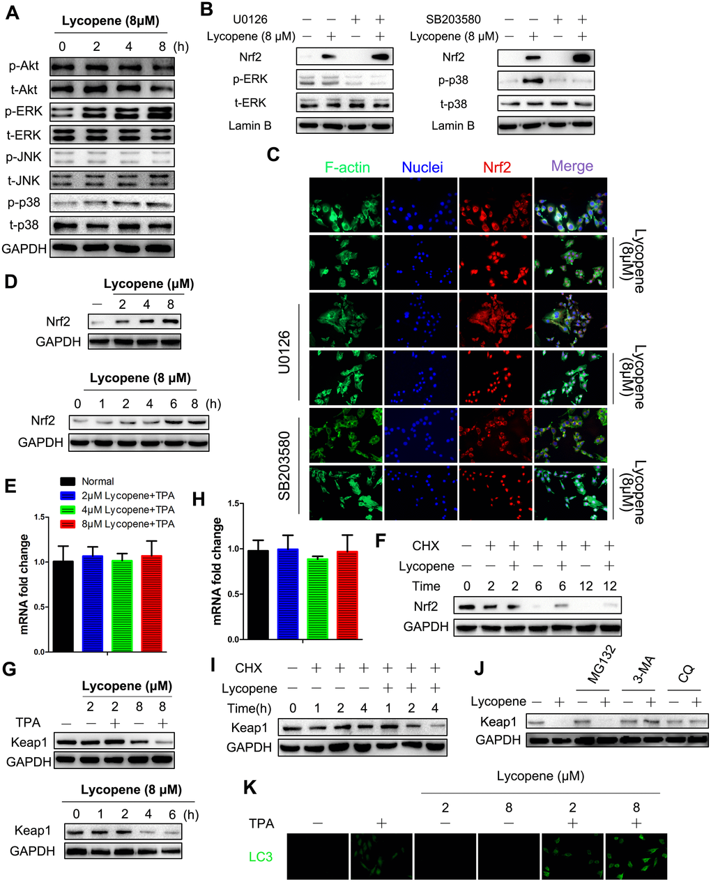
Figure 7.Lycopene induced activation of Nrf2 by reducing Keap1 protein at the posttranslational level via the autophagy-lysosomal pathway. (A) Cells were incubated with 8 μΜ lycopene for the indicated time and then exposed with TPA for additional 2 hours, and the expression of both phosphorylated and total forms of Akt, ERK1/2, p38 and JNK1/2 were measured by western blot analysis. (B) Cells were pre-treated with U0126 (left) or SB203580 (right), followed by lycopene treatment for 8 h. The nuclear protein extract was subjected to immunoblot analysis for the detection of Nrf2 expression. (C) Immunofluorescence analysis of Nrf2 was carried out as described in Methods. Treatment was similar to (B). (D) Time-dependent (bottom) and dose-dependent (top) study of lycopene on Nrf2 protein levels. Cells were pretreated with 8 lycopene for different times or with different doses of lycopene for 12 h and then exposed with TPA for additional 2 hours, and the Nrf2 protein level was assayed by Western blot. (E) Total RNA was isolated and analyzed to determine the levels of Nfe2l2 mRNA using real-time qPCR after lycopene treatment for 12 hours. House-keeping gene gaphd was used as the internal control. The data are presented as the mean ± SD. (n=3). (F) Cells were pretreated with CHX (0.5 g/ml) alone or in the presence of lycopene (8 μM) and TPA for various times. Nrf2 protein was examined by Western blot. (G) Time-dependent (right) and dose-dependent (left) study of lycopene on keap1 protein levels. Cells were pretreated with 8 μM lycopene for different times or with different doses of lycopene for 4 h and then exposed with TPA for additional 2 hours, and the keap1 protein level was assayed by western blot. (H) Total RNA was isolated and analyzed to determine the levels of Keap1 expression using real-time qPCR after lycopene treatment for 6 hours. House-keeping gene gapdh was used as the control. The data are presented as the mean ± SD. (n=3). (I) Cells were pretreated with CHX (0.5 g/ml) alone or in the presence of lycopene (8 μM) and TPA for various times. keap1 protein was examined by Western blot. (J) Cells were treated with MG132 (1 μM) or 3-MA (1 mM) or CQ (25 μM) for 1 h. Lycopene (8 μM) was added to cells for 6 hours, and then exposed with TPA for additional 2 hours. Expression of keap1 protein was examined by western blot. (K) Dose-dependent study of lycopene on LC3 protein levels. Cells were pretreated with different doses of lycopene for 6 h and then exposed with or without TPA for additional 2 hours, and the LC3 was analyzed by immunofluorescent staining. The results are representative blot images of three independent experiments in A, B, D, F, G, I, J, respectively.