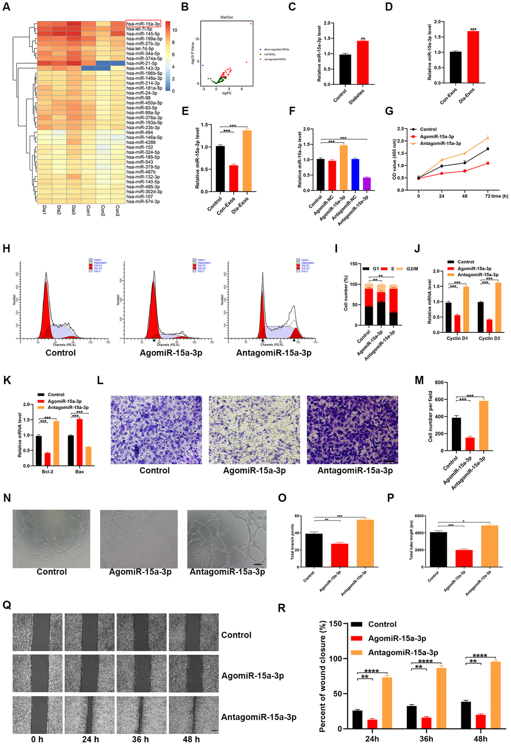
Figure 4.Dia-Exos were enriched with miR-15a-3p, which altered HUVEC function. (A, B) An miRNA microarray dataset of non-diabetic foot wound patients and DFU patients retrieved from NCBI GEO (accession number: GSE80178) indicated that miR-15a-3p was upregulated in foot skin from diabetic patients. (C, D) MiR-15a-3p overexpression was found in serum and exosomes from the diabetic group; n = 10 per group. (E) Effects of the two kinds of exosomes on miR-15a-3p levels in the skin tissues of mice treated with Dia-Exos. (F) qRT-PCR indicated that antagomiR-15a-3p could partially counteract the overexpression of miR-15a-3p in HUVECs. (G) A CCK-8 assay was used to assess the effects of antagomiR-15a-3p on HUVEC proliferation. (H, I) Flow cytometry was used to quantify the cell cycle distribution. (J) qRT-PCR analysis indicated that antagomiR-15a-3p could restore the mRNA levels of Cyclin D1 and Cyclin D3. (K) The effects of antagomiR-15a-3p on the apoptosis-related genes Bcl-2 and Bax were measured using qRT-PCR. (L, M) A Transwell migration assay was used to measure the effects of miR-15a-3p on HUVEC migration; scale bar: 100 μm. (N–P) A tube formation assay was used to assess the effects of miR-15a-3p on HUVEC angiogenesis; scale bar: 200 μm. (Q, R) The scratch assay results of the three groups; scale bar: 250 μm. Data are the means ± SDs of three independent experiments. *p < 0.05, **p < 0.01, ***p < 0.001.