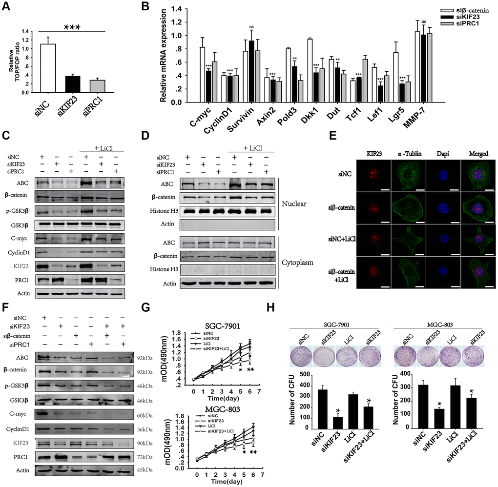
Figure 3.Loss of KIF23 impaired the Wnt/β-catenin signaling pathway in GC. (A) TCF luciferase reporter plasmid and its mutant plasmid were constructed and transfected into MGC-803 cells. Changes in endogenous Wnt/TCF reporter activity in MGC-803 cells after silencing of KIF23 or PRC1 were analyzed. (B) Q-PCR analysis of the effects of KIF23 and PRC1 silencing on 11 Wnt target genes in MGC-803 cells. (C) Western blot analysis of the levels of KIF23, β-catenin activation and Wnt targets in MGC-803 cells treated with siNC, KIF23 siRNA (siKIF23) or PRC1 siRNA (siPRC1) with or without LiCl for activation of the Wnt/β-catenin signaling pathway overnight. (D) Nuclear and plasma proteins were extracted, and Western blot analysis of β-catenin activation was performed to reveal the distribution of β-catenin in nuclear and cytoplasmic fractions (Histone H3: nuclear protein marker, actin: cytoplasmic protein marker). (E) Immunofluorescence staining for nuclear β-catenin after siRNAs application. β-catenin was labeled in red, and the cytoskeleton protein was labeled in green. (F) Western blot analysis of β-catenin activation and Wnt targets after rescue of PRC1 levels with siRPC1. Actin was used as the loading control. Cell growth (G) and colony formation assays (H) were performed after KIF23 knockdown and LiCl activation. All experiments were repeated at least three times. Statistically significant differences are indicated.* p < 0.05, ** p < 0.01, *** p < 0.001; ns, no significant difference.