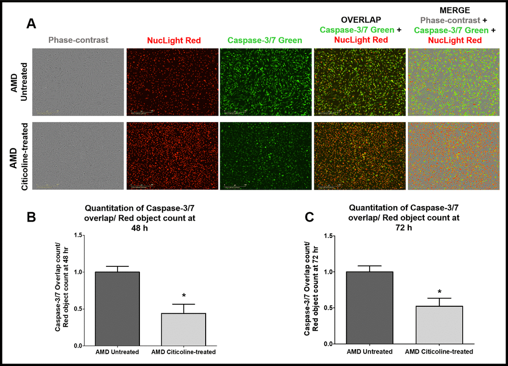
Figure 3.(A) Upper and lower panels show Representative Incucyte live-cell images of untreated and Citicoline-treated AMD cells ,respectively, in phase-contrast (first column), stained with NucLight Red (second column), stained with Caspase-3/7 Green (third column), overlap i.e., Caspase-3/7 + NucLight (fourth column), and Merge i.e., Phase-contrast + Caspase-3/7 + NucLight (fifth column). Scale bar = 400 μM.(B) Quantitation of Caspase-3/7 overlap/ Red object count at the 48 h time point. (C) Quantitation of Caspase-3/7 overlap/ Red object count at the 72 h time point.