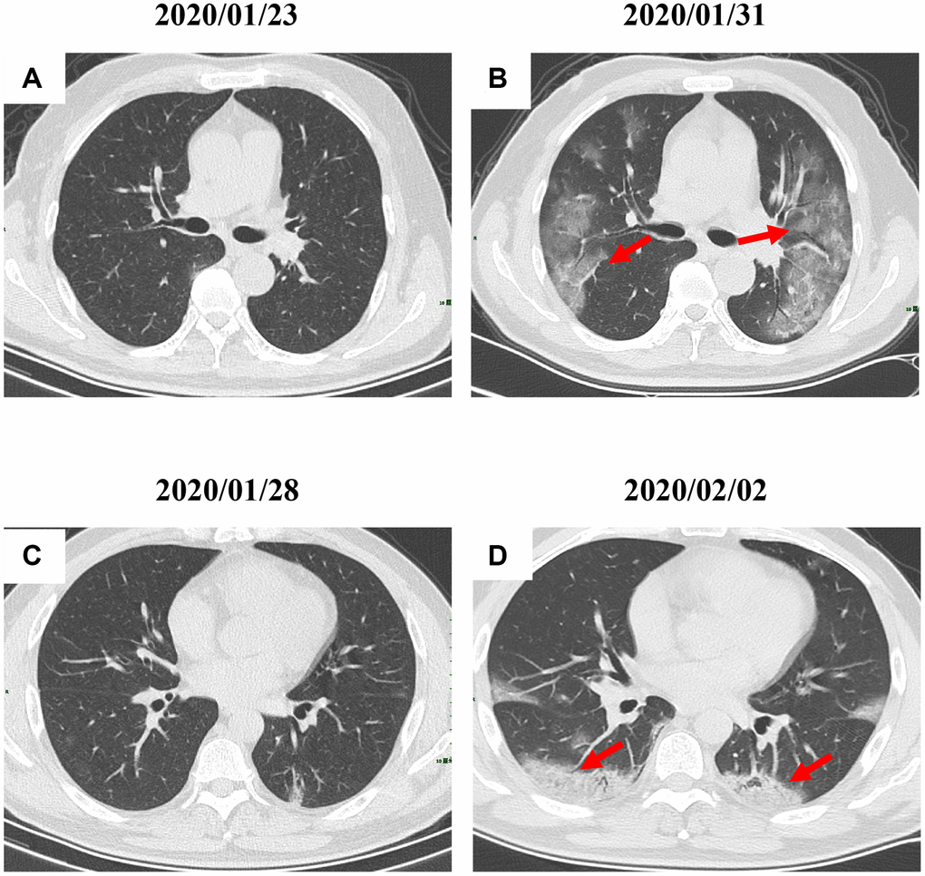
Figure 3.Radiological worsen progression of two COVID-19 pneumonia patients. (A, B): Bilateral, large, and multiple ground-glass opacity was observed in a 47-year-old male patient with type 2 diabetes after 8 days since admission; (C, D) Consolidation accompanying air bronchogram were found in the bilateral lower lungs of a 29-year-old male patient after 5 days since admission. Typical lesions were marked with red arrows.