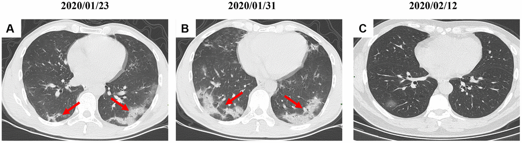
Figure 6.A serial CT images after admission of a 54-year-old female patient. (A) Patch ground-glass opacity mainly located in the left lower lobe. (B) Significant larger patch ground-glass opacities were observed in both lower lobes after 8 days. (C) Follow-up CT scans on day 20 after admission show a remarkable improvement. Typical lesions were marked with red arrows.