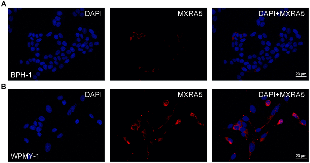
Figure 4.Immunofluorescence of MXRA5 in human prostate cells. (A) Human epithelial cells (BPH-1). Left: DAPI (blue) indicates nuclear staining. Middle: Cy3-immunofluorescence (red) indicates the MXRA5 protein which was rarely observed in the epithelial cells. Right: Merged image. The scale bar is 20 μm. (B) Human stromal cells (WPMY-1). Left: DAPI (blue) indicates nuclear staining. Middle: Cy3-immunofluorescence (red) indicates the MXRA5 protein which was abundantly observed in the stromal cells. Right: Merged image. The scale bar is 20 μm. Representative graphs of prostate cells were selected into figure.