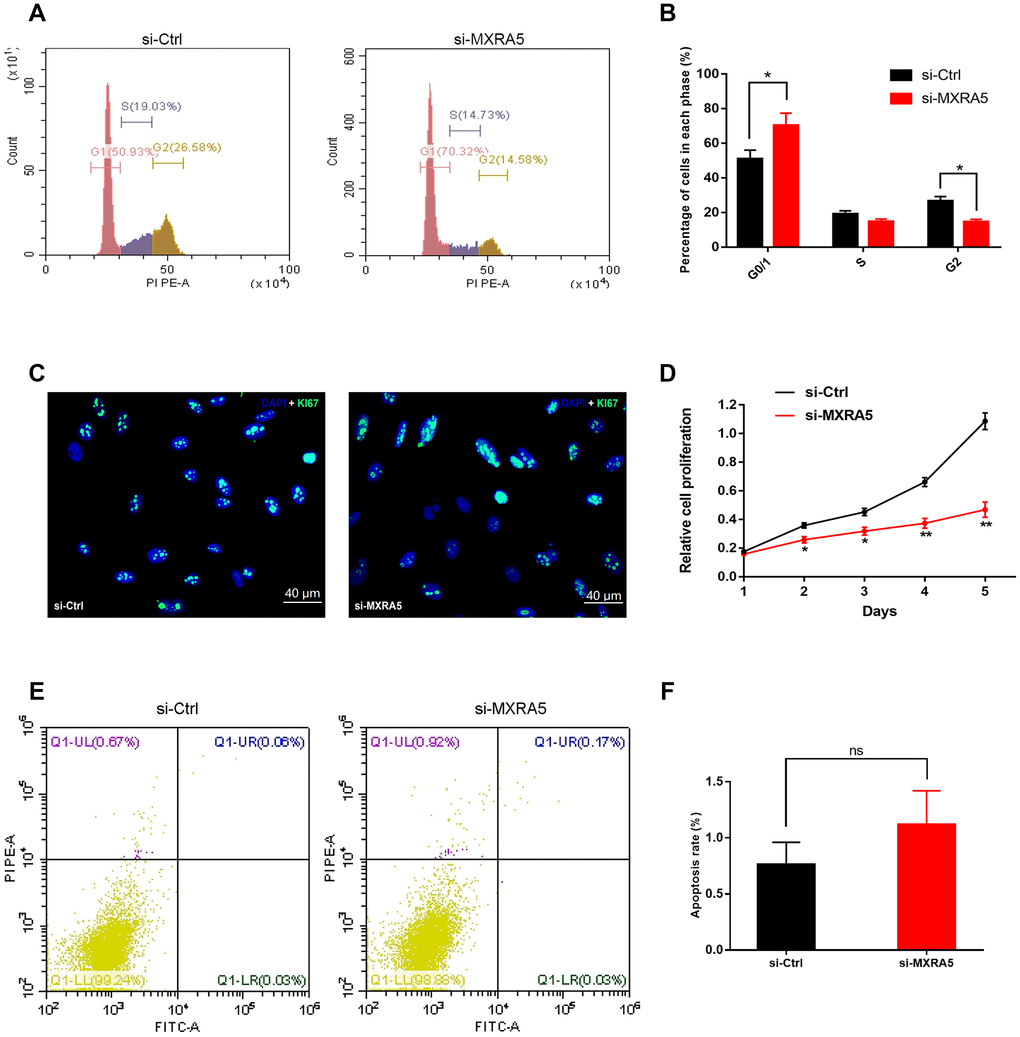
Figure 6.Downregulation of MXRA5 represses prostate cell proliferation. (A, B) Flow cytometry analysis for the WPMY-1 cells treated with si-MXRA5 for 48h compared with si-control treated cells. Percentages (%) of cell populations at different stages of cell cycles were listed within the panels. All histograms revealed the percentage (%) of cell populations from three independent experiments, * means P < 0.05. (C) Cell proliferation of WPMY-1 cells treated by si-control and si-MXRA5 was detected by Ki-67 staining (green). Nuclei were stained by DAPI (blue). Three repeats of experiments for were conducted and representative graphs were selected into figure. The scale bar is 40 μm. (D) MTT assay was used to detect the viability of the WPMY-1 cells treated by si-control (black line) and si-MXRA5 (red line). (E) Flow cytometry analysis of alterations of WPMY-1 cells apoptosis by the transfection using si-control and si-MXRA5. PI PE-A in y-axis stands for the fluorescence intensity of propidine iodide (PI) and FITC-A in x-axis stands for the fluorescence intensity of Fluorescein isothiocyanate (FITC) laballed Annexin V. Calculation area of the apoptosis rate was percentage of Annexin V+/PI+ cells. (F) Statistical analysis suggested no significant (ns) induction of apoptosis by the downregulation of MXRA5 in WPMY-1 cells. All values shown are mean ± SD of triplicate measurements and repeated three times with similar results, * means P < 0.05 and ** means P < 0.01.