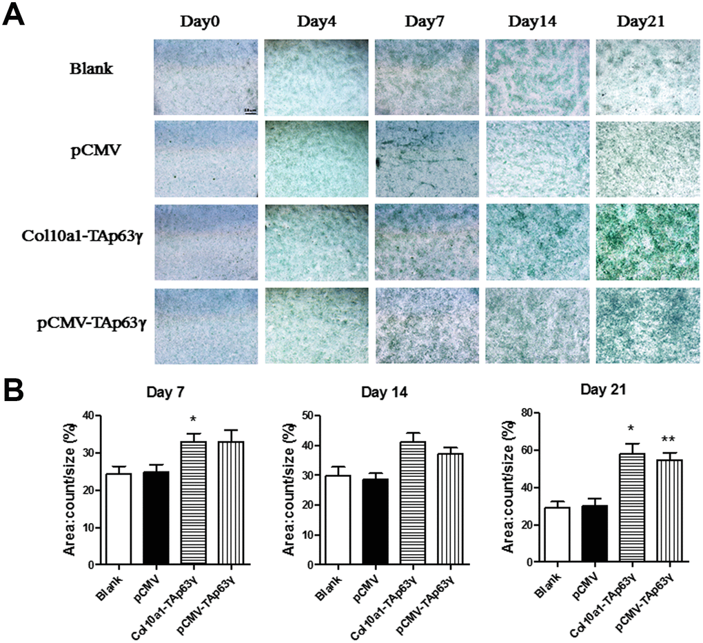
Figure 3.In vitro effect of TAp63γ on chondrocyte proliferation. (A) ATD5C cells were cultured for 0, 4, 7, 14, or 21 days and stained with Alcian blue. After 7 days in culture, the staining intensity of TAp63γ stable cell lines was much stronger than the blank and vector controls. Scale bar, 25 μm. (B) Sum object area of the staining by densitometry analysis (n=3, * p<0.05, ** p<0.01).