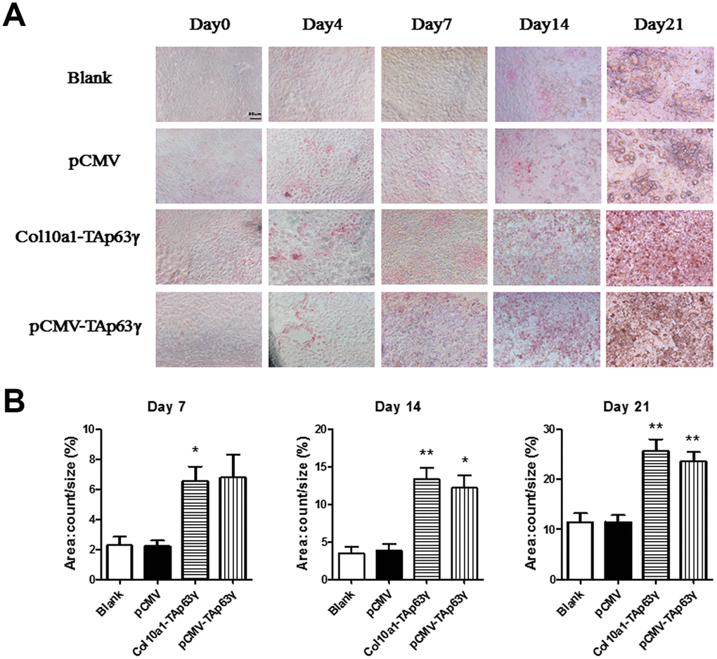
Figure 4.TAp63γ promotes hypertrophic differentiation of ATDC5 cells. (A) ATD5C cells were cultured for 0, 4, 7, 14, or 21 days and stained for ALP (alkaline phosphatase). After 7 days in culture, the staining intensity of TAp63γ stable cell lines was much stronger than the blank and vector controls. Scale bar, 50 μm. (B) Sum object area of the staining by densitometry analysis (n=3, * p<0.05, ** p<0.01).