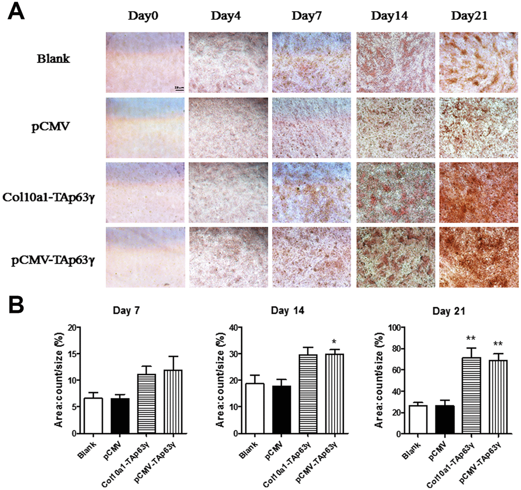
Figure 5.In vitro effect of TAp63γ on matrix mineralization. (A) ATD5C cells were cultured for 0, 4, 7, 14, or 21 days and stained with Alizarin red. After 4 and 7 days in culture, enhanced Alizarin red staining was observed in both Col10a1-TAp63γ and pCMV-TAp63γ stable cell lines compared with the blank and vector controls. Scale bar, 25 μm. (B) Sum object area of the staining by densitometry analysis (n=3, * p<0.05, ** p<0.01).