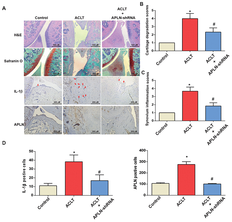
Figure 6.shAPLN administration mitigates the histologic severity of OA. (A) Staining of specimens with H&E, Safranin-O, IL-1β and APLN from the control knee (n=6), ACLT knee (n=6), and shAPLN-transfected ACLT knee (n=6). (B) Cartilage degeneration scores were calculated for articular cartilage sections stained with Safranin-O. (C) Synovial membrane inflammation score. Magnified area of synovium used to generate synovial inflammation score in all samples. Scoring was performed in H&E-stained slides. (D) IHC analysis of proportions of IL-1β-positive cells (red arrows) and APLN-positive cells in synovial lining tissues in specimens from control knees (n=6), ACLT knees (n=6), and shAPLN-transfected ACLT knees (n=6). * p<0.05 compared with the control group; # p<0.05 compared with the control shRNA-transfected ACLT group.