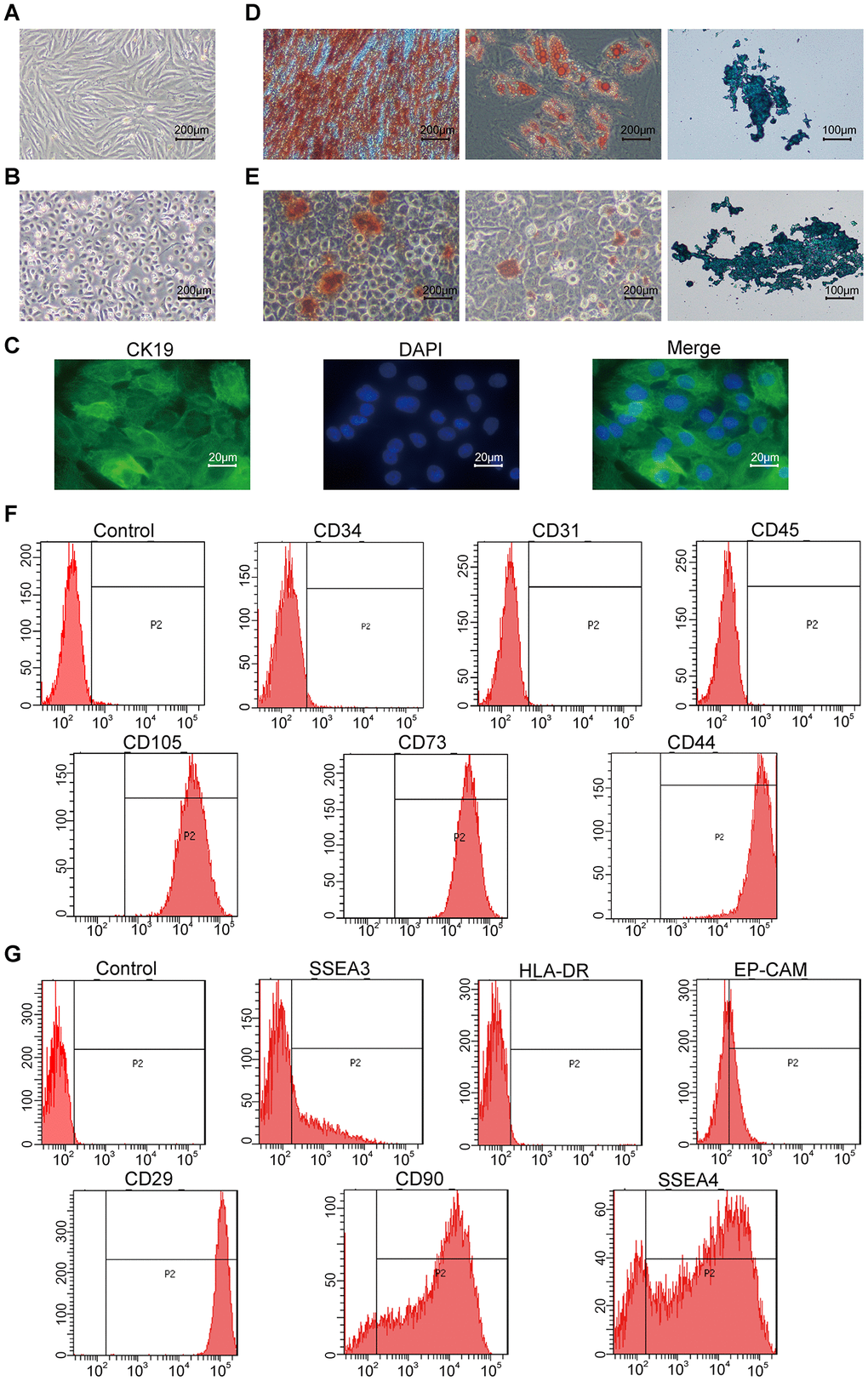
Figure 1.Characterization of hAMSCs (Human amniotic mesenchymal stem cells) and hAECs (Human amniotic epithelial cells). (A, B) Representative phase-contrast bright field images (scale bar: 200 μm) show confluent cultures of (A) hAMSCs and (B) hAECs. (C) Fluorescence images (scale bar: 20 μm) show positive expression of the epithelial stem cell marker Cytokeratin 19 (CK19; green) on the keratinocytes. The nuclei are stained with DAPI (blue). (D, E) Representative images show alizarin red (scale bar: 200 μm), alcian blue (scale bar: 200 μm), and oil red O (scale bar: 100 μm) stained hAMSCs (D) and hAECs (E) that have undergone osteogenic adipogenic or chondrogenic differentiation, respectively. (F) Flow cytometry analysis shows surface expression of CD34, CD31, CD45, CD105, CD73, and CD44 on the hAMSCs. (G) Flow cytometry analysis shows surface expression of SSEA3, HLA-DR, Ep-CAM, CD29, CD90, and SSEA4 on hAMCs.