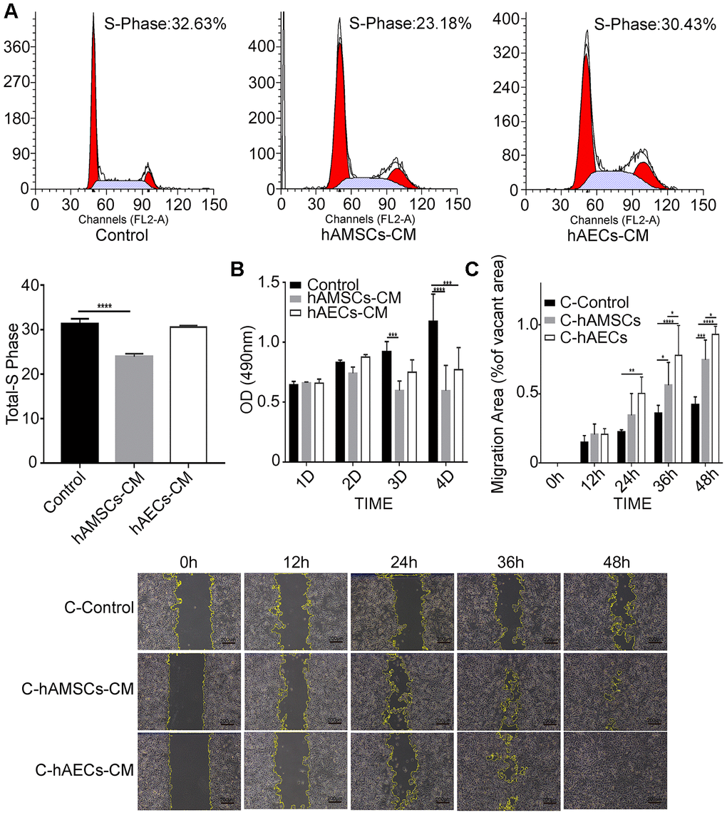
Figure 3.In vitro culturing with hAMSCs-CM (condition media of hAMSCs) and hAECs-CM (condition media of hAECs) inhibits proliferation, but promotes migration of keratinocytes. (A) FACS plots show cell cycle analysis of keratinocytes grown in control medium, hAMSCs-CM and hAECs-CM. The cells were stained with propidium iodide. Histogram shows the percentage of S-phase keratinocytes when grown in control medium, hAMSCs-CM and hAECs-CM. (B) Histogram plot shows analysis of keratinocyte proliferation in control medium, hAMSCs-CM and hAECs-CM on days 1, 2 and 3, and 4 using the MTS assay. (C) Histogram plot shows results of the scratch wound assay. (Top) The cell migration area is plotted for each group of keratinocytes at various time points (0, 12, 24, 36, and 48 h). The phase contrast bright field images (Bottom) show the status of keratinocyte migration in the control medium, hAMSCs-CM and hAECs-CM. The cells were pretreated with mitomycin C to normalize differences in proliferation. Note: The values are expressed as means ±SEM. ****p < 0.0001; ***p < 0.001; **p < 0.01; *p < 0.05.