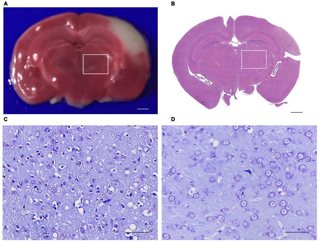
Figure 1.Triphenyltetrazolium chloride (TTC), Hematoxylin and eosin (H&E) and Nissl Staining of the brain section. (A) TTC staining showed the red region is normal brain tissue and the white region is infarction (bar=100μm). (B) H&E staining showed that the primary infarction is confined to the left cortex and does not involve the ipsilateral thalamus in mice (bar=100μm). (C, D) Nissling staining of the ipsilateral thalumus of the focal cortical infarction mice and sham mice (bar=50μm). (The rectangle indicates the non-ischemic ipsilateral thalamus).