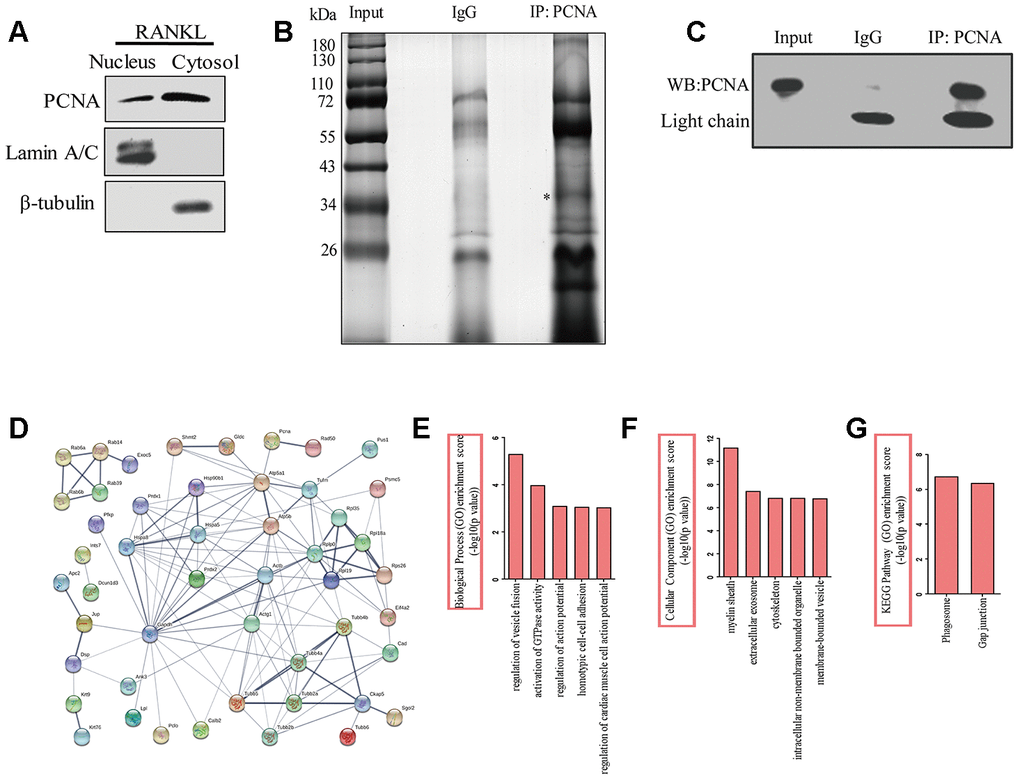
Figure 4.The identification of the proteins interacting with cytoplasmic PCNA. (A) RAW264.7 cells after RANKL (100 ng/mL) induction for three days were subjected to fractionation experiments to isolate the nuclear and cytoplasmic fractions. The distribution of PCNA in both fractions was assessed by western blotting. Lamin A/C and β-tubulin were respectively used as markers for nuclear and cytoplasmic fractions. (B) Co-IP assay using PCNA antibody was carried out with the cytoplasmic fraction, PCNA-bound proteins were separated using SDS-PAGE gel electrophoresis and silver stained. IP with IgG was used as the control. The asterisk denotes the protein band corresponding probably to the immunoprecipitated PCNA protein. (C) Confirmation by western blotting of the successful immunoprecipitation of PCNA in (B). Light chain: the light chain of IgG and PCNA antibody. (D) STRING program analysis of the interaction network of 76 putative cytoplasmic PCNA interactors. Line thickness indicates the strength of data support. (E) Biological processes analysis of the identified differential proteins using the ClueGO plug-in of Cytoscape software. The top five items were listed. (F) Cellular component enrichment analysis of the identified differential proteins using Cytoscape software. The top five items were listed. (G) KEGG pathway enrichment analysis of the identified differential proteins using Cytoscape software. The top two items were listed.