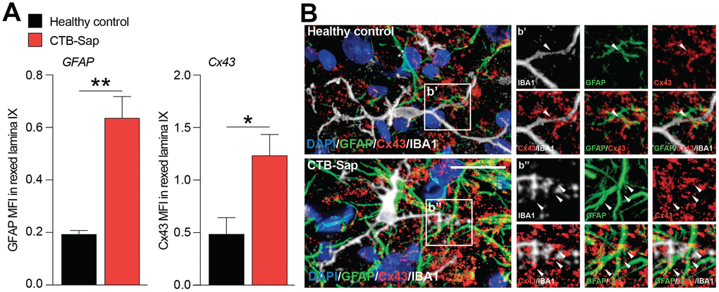
Figure 4.Increase of Cx43 in the spinal cord cell populations of motoneuron-depleted spinal cord. (A) Quantification of mean fluorescence intensity (MFI) of GFAP and Cx43 in the left lamina IX of healthy control and CTB-Sap lesioned mice; data are expressed as mean ± SEM; **p-value < 0.01 and *p-value < 0.05 versus healthy control. (B) Representative confocal images of Cx43 (red) immunofluorescence analysis in lamina IX of healthy control and CTB-Sap lesioned mice; images show also markers for astroglial cells (GFAP, green) and microglia (IBA1, white); scale bar 20 μm.