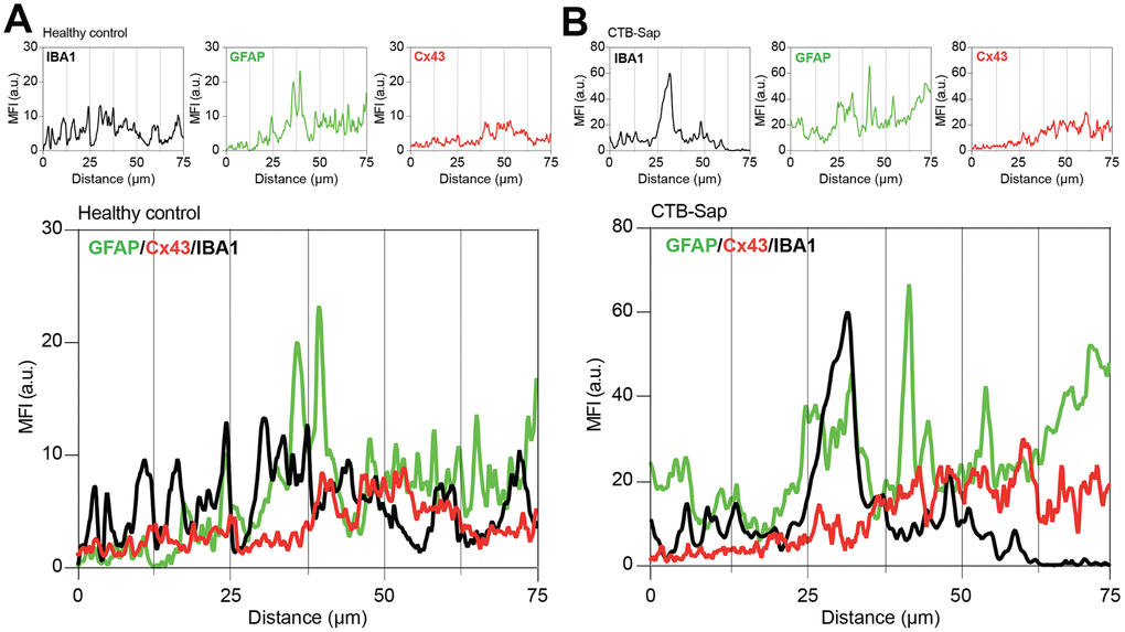
Figure 5.Cx43-based channels profile in microglial/astroglial milieu in motoneuron-depleted spinal cord. Profile plot of MFI of IBA1 (black plot), GFAP (green plot), and Cx43 (red plot) and plots overlay (bottom panel) in Rexed lamina IX of healthy control (A) and CTB-Sap lesioned mice (B); data are MFI arbitrary units (a.u.) of spinal confocal acquisitions.