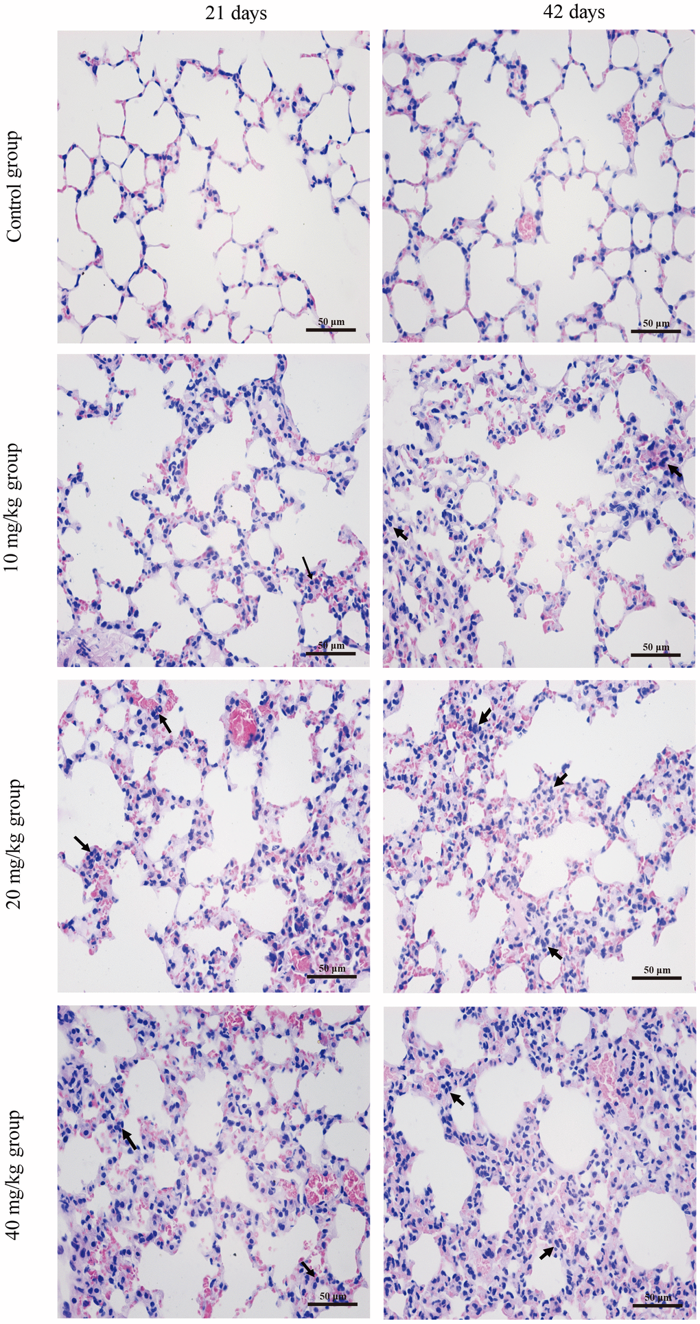
Figure 1.Histopathological changes in the lung at 21 and 42 days of the experiment. (H&E ×400). Control group at 21 and 42 days: no changes are observed. 10 mg/kg group at 21 days: the inflammatory cells (↑) are obviously observed in alveolar walls. 20 mg/kg group at 21 days: the alveolar walls are slightly thickened with inflammatory cell infiltration (↑) 40 mg/kg group at 21 days: the alveolar walls are obviously thickened with inflammatory cell infiltration (↑). 10 mg/kg group at 42 days: the alveolar walls are slightly thickened with inflammatory cell infiltration (↑). 20 mg/kg group at 42 days: the alveolar walls are thickened with inflammatory cell infiltration (↑). 40 mg/kg group at 42 days: the alveolar walls are markedly thickened with inflammatory cell infiltration (↑).