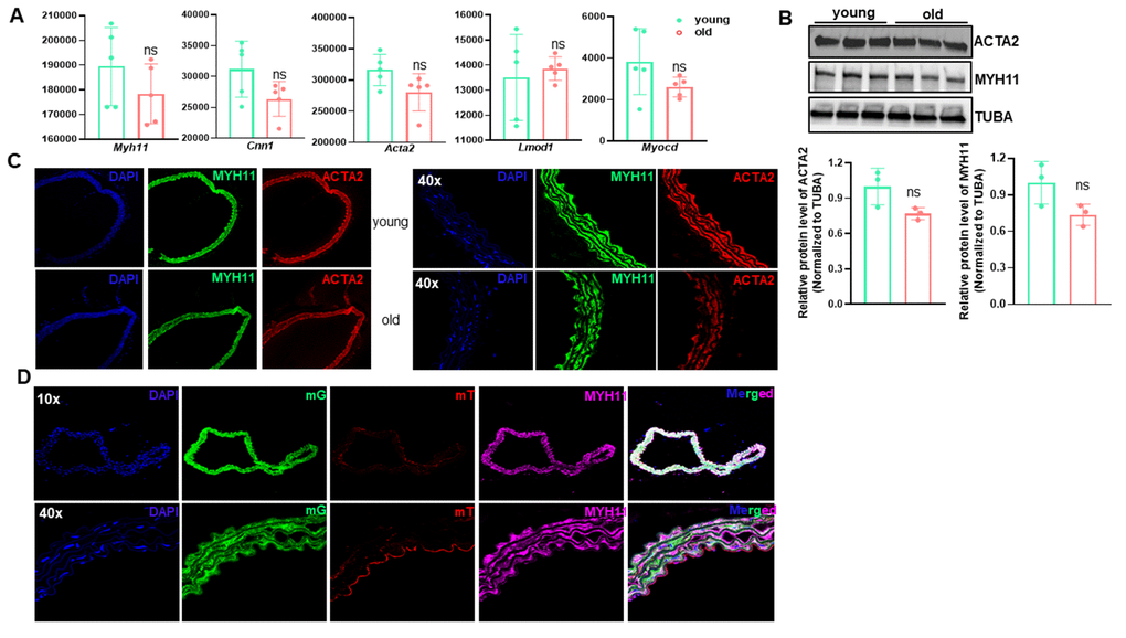
Figure 7.The contractile vascular smooth muscle cell (VSMC) phenotype is retained in old aortae. (A) Normalized counts of SMC marker genes in young and old mouse aortae. Data are presented as a scatterplot of individual points with mean±SD, n=5. ns, not significant compared to young aortae, unpaired two-tailed Student's t-test. (B) Western blotting for the indicated SMC marker proteins in total protein lysates of young and old mouse aortae and its quantitation (n=3). (C) Representative confocal microscopy images of immunofluorescence staining for MYH11 and ACTA2 in aortae of young and old C57/BL6 mice. (D) Representative confocal microscopy images of immunofluorescence staining for MYH11 and ACTA2 together with fluorescence of membrane Tomato (mT, Red) and membrane GFP (mG, Green) in young and old Myh11-Cre-ERT/mTmG reporter mice. Mice (14 wks) were injected with tamoxifen (TMX) for 5 consecutive days (n=4) and aortae were isolated at the age of 54 wks. Upon TMX induction, all mature SMCs were labeled by mG, whereas other cell types were labeled by mT.