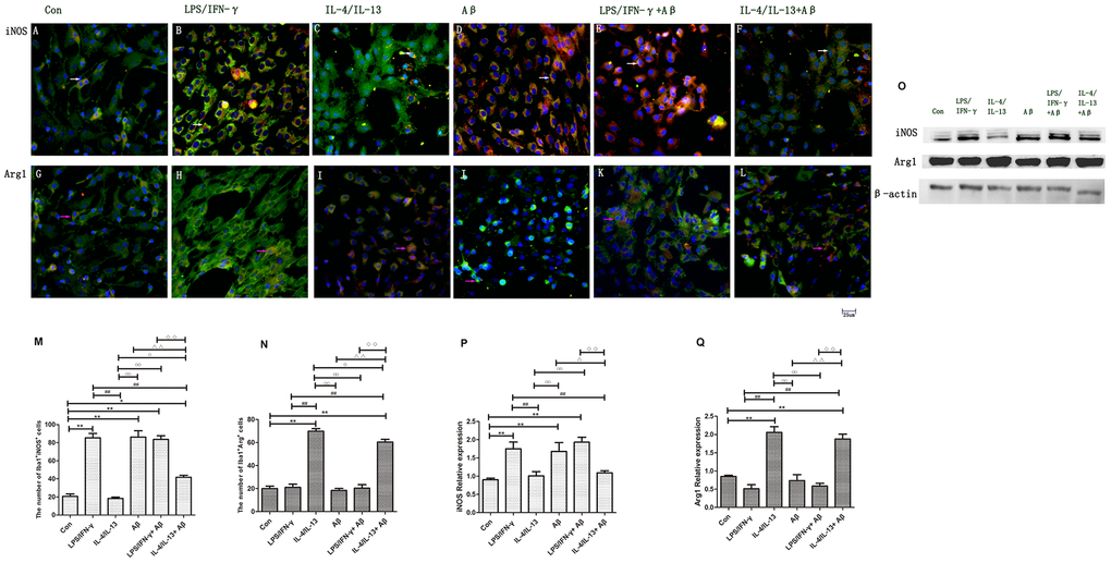
Figure 2.Microglial polarization was induced by inflammatory factors and Aβ. (A–F) Microglia in different groups were double-stained with anti-iNOS and anti-Iba1 antibodies and observed on a fluorescence microscope. Green represents Iba1+ cells, red represents iNOS+ cells and blue represents DAPI staining. The white arrows denote iNOS+Iba1+ cells. (G–L) Microglia in different groups were double-stained with anti-Arg1 and anti-Iba1 antibodies. Green represents Iba1+ cells, red represents Arg1+ cells and blue represents DAPI staining. White arrows denote Arg1+Iba1+ cells. Scale bars, 25 μm. (M, N) Quantitative data on the mean number of iNOS+Iba1+ or Arg1+Iba1+ cells (n=5). (O) Microglia in different groups were subjected to Western blotting to detect iNOS and Arg1. β-Actin was used as the internal control. (P, Q) Quantitative data on the relative protein levels of iNOS and Arg1 (n=3). Error bars, S.E.M. Compared with Con, *p<0.05, **p<0.01. Compared with LPS/IFN-γ, #p<0.05, ##p<0.01. Compared with IL-4/IL-13, ○p<0.05, ○○p<0.01. Compared with Aβ, Δp<0.05, ΔΔp<0.01. Compared with LPS/IFN-γ+Aβ, ↓p<0.05, ↓↓↓p<0.01. One-way ANOVA was performed with Tukey’s correction.