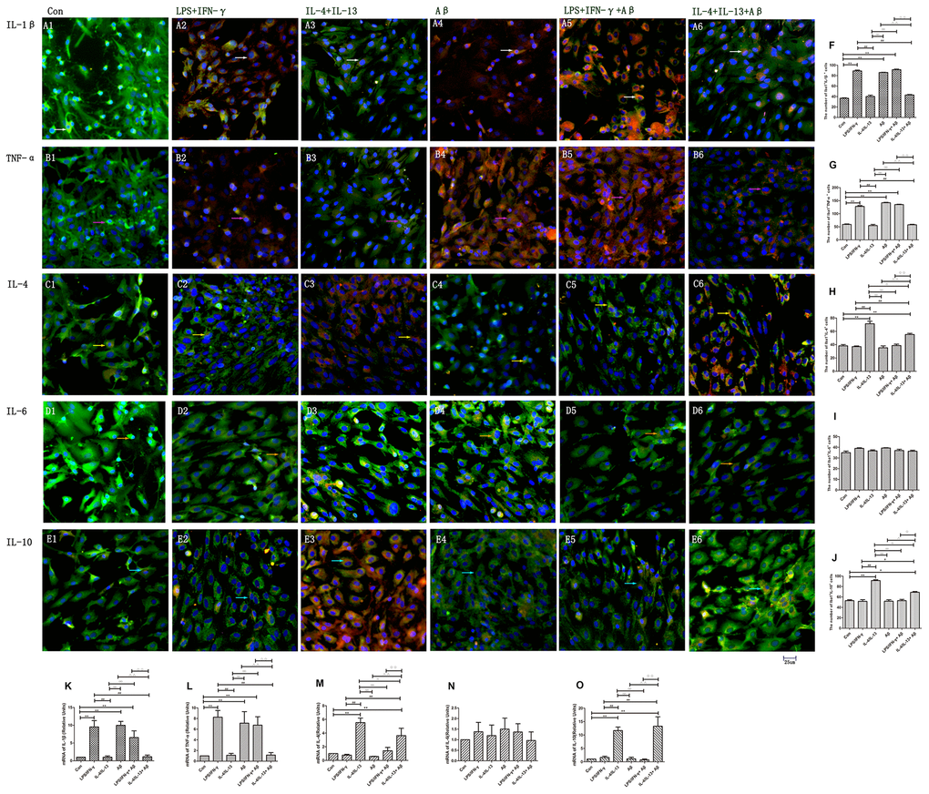
Figure 3.Microglia produced various cytokines under different stimuli. (A–E) Microglia in different groups were double-stained with antibodies against various cytokines and Iba1. (A1–A6) Double-staining for IL-1β and Iba1. (B1–B6) TNF-α and Iba1. (C1–C6) IL-4 and Iba1. (D1–D6) IL-6 and Iba1. (E1–E6) IL-10 and Iba1. Green represents Iba1+ cells, red represents cytokine-positive cells and blue represents DAPI staining. White arrows denote IL-1β+Iba1+ cells. Pink arrows denote TNF-α+Iba1+ cells. Yellow arrows denote IL-4+Iba1+ cells. Beige arrows denote IL-6+Iba1+ cells. Blue arrows denote IL-10+Iba1+ cells. (F–J) The numbers of double-positive cells in different groups (n=3). (K–O) Quantitative data on the relative mRNA levels of cytokines in microglia (n=3). Error bars, S.E.M. Compared with Con, *p<0.05, **p<0.01. Compared with LPS/IFN-γ, #p<0.05, ##p<0.01. Compared with IL-4/IL-13, ○p<0.05, ○○p<0.01. Compared with Aβ, Δp<0.05, ΔΔp<0.01. Compared with LPS/IFN-γ+Aβ, ↓p<0.05, ↓↓p<0.01. One-way ANOVA was performed with Tukey’s correction.