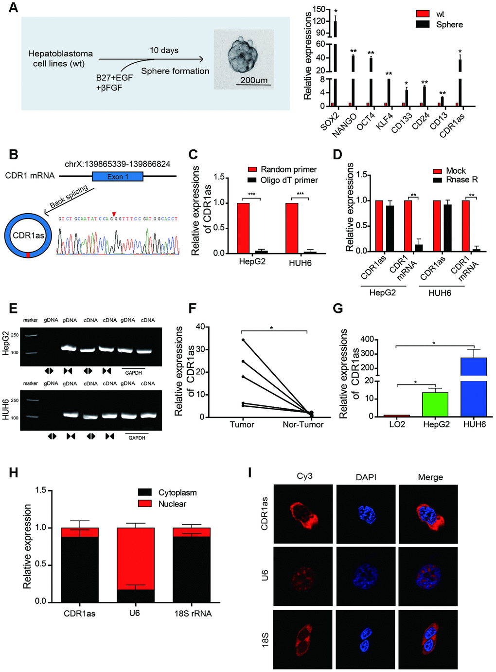
Figure 1.The identification, characteristics, and expression of CDR1as in HB cell lines and HB. (A) Schematic illustration of a forming HB sphere and qRT-PCR analysis for cancer stem cell (CSC) markers; (B) The formation of CDR1as. CDR1as is derived from back-spliced exon 1 of genomic CDR1. The existence of CDR1as was confirmed by Sanger sequencing. The red arrow represents the back-splice junction of CDR1as; (C) qRT-PCR analysis of CDR1as: reverse transcription products using random primers or oligo dT primers; (D) The expression of CDR1as and CDR1 mRNA was measured by qRT-PCR in HepG2 and HUH6 cells that were treated with or without RNase R; (E) The PCR products of CDR1as and linear CDR1 were evaluated by gel electrophoresis. Divergent primers amplified CDR1as in cDNA but not genomic DNA (gDNA). Convergent primers amplified linear CDR1 in both cDNA and gDNA. GAPDH was used as a linear control; (F) qRT-PCR analysis of CDR1as in 5 paired HB and adjacent noncancerous tissues; (G) The expression of CDR1as in HB cell lines was detected by qRT-PCR; (H) CDR1as was mainly located in the cytoplasm as confirmed by the nuclear mass separation assay in HepG2 cells; (I) Fluorescence in situ hybridization (FISH) confirmed that CDR1as was predominantly located in the cytoplasm. Nuclei were stained with DAPI. U6, 18S, and CDR1as were labeled with Cy3. Scale bar, 200 μm. Data are presented as the mean ± SEM of three experiments. *P < 0.05,**P < 0.01,***P < 0.001 (Student’s t-test).