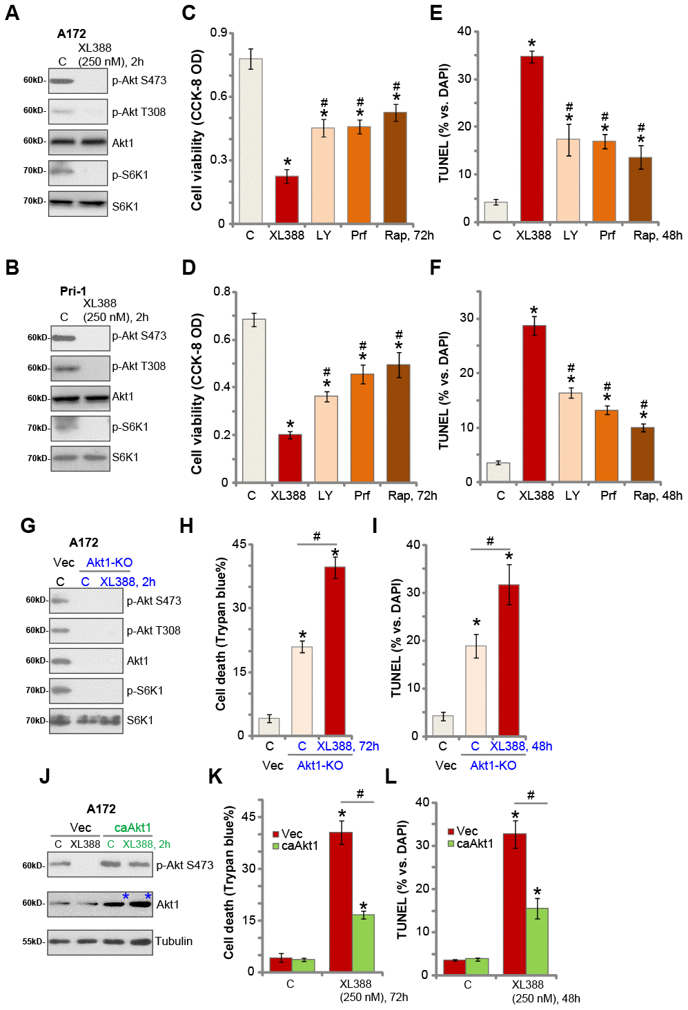
Figure 3.XL388-induced anti-glioma cell activity is through Akt-mTOR-dependent and -independent mechanisms. A172 cells or the primary human glioma cells, Pri-1, were treated with XL388 (250 nM), and cultured for 2h, and expression of listed proteins was shown (A and B). A172 cells or the Pri-1 primary human glioma cells were treated with XL388 (250 nM), LY294002 (“LY”, 1 μM), perifosine (“Prf”, 5 μM) or rapamycin (“Rap”, 500 nM) for 48-72h, then cell viability and apoptosis were tested by CCK-8 (C and D) and TUNEL staining (E and F) assays, respectively. Stable A172 cells with the CRISPR/Cas9-Akt1-KO construct (“Akt1-KO” cells) or empty vector (“Vec”) were treated with or without XL388 (250 nM) for applied time, and cultured for applied time periods, and expression of listed proteins was shown (G);Cell death and apoptosis were tested by Trypan blue staining (H) and nuclear TUNEL staining (I) assays, respectively. Stable A172 cells with a constitutive-active Akt1 (S473D, “ca-Akt1”) or empty vector (“Vec”) were treated with or without XL388 (250 nM) for applied time, and expression of listed proteins was shown (J); cell death and apoptosis were tested by Trypan blue staining (K) and nuclear TUNEL staining (L) assays, respectively. Data were presented as mean ± SD (n=5).* p <0.05 vs. “C” cells. #p <0.05 vs. XL388 treatment (C–F). #p <0.05 (H, I, K and L). Experiments in this figure were repeated three times, and similar results were obtained.