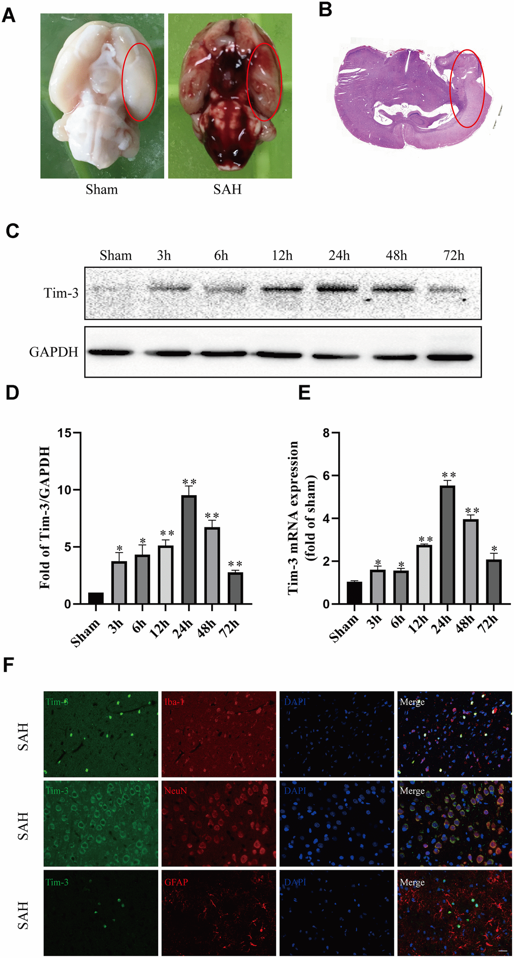
Figure 1.Representative image of the subarachnoid hemorrhage (SAH) model and endogenous expression of Tim-3 in brain tissue. Representative images of brains from the sham and SAH groups. (A) A schematic indicating the optimal brain region for immunochemical staining, qRT-PCR, and western blotting (red circle). (B) Western blot analysis showed Tim-3 protein abundance at 3, 6, 12, 24, 48, and 72 h after SAH. (C) Quantification of the Tim-3 protein level. (D) Quantification of Tim-3 mRNA level in the rat brain. (E) Representative microphotographs of immunofluorescence staining for Tim-3 and Iba1, NeuN, GFAP. (F) All values are presented as means ± SD, n = 6 for each time point per group. *p < 0.05, **p < 0.01 versus sham group. Scale bar = 50 μm.