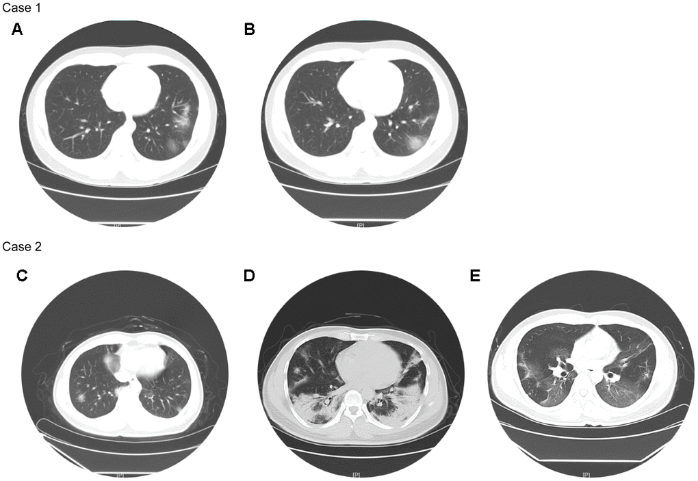
Figure 1.Chest CTs of two representative cases. Case 1 (non-severe): Chest CT on Feb 24 (A) showed multiple patchy ground-glass opacity in both lungs, with unclear borders and uneven density. Chest CT on Feb 28 (B) showed better status, and some lesions were slightly absorbed than before. Case 2 (severe): Chest CT on Jan 29 (C) showed the texture of both lungs was slightly increased, and both lungs were scattered in patchy shadows, whose edges were blurred. Chest CT on Feb 11 (D) showed the scope of the bilateral lung lesions was enlarged, the density was increased, and the local consolidation and bronchial signs were seen. Chest CT on Mar 4 (E) showed improved status, and both lung lesions were significantly less than before.