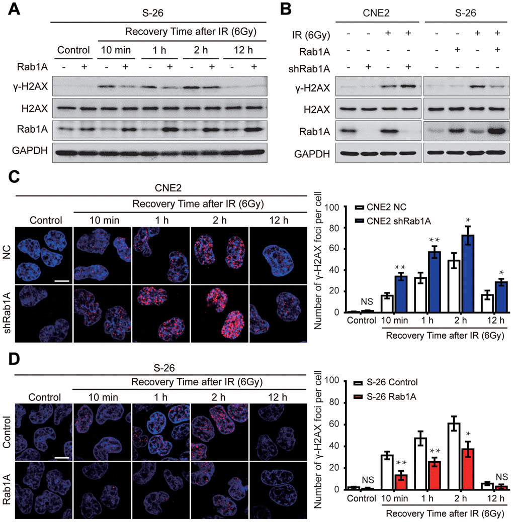
Figure 4.Rab1A inhibits IR-inducible phosphorylation of serine 139 of H2AX (γ-H2AX) in NPC cells. (A) Immunoblot analysis of 6 Gy IR-induced time-dependent changes of γ-H2AX, H2AX and Rab1A. Non-irradiated or irradiated S-26 cells with Rab1A overexpression or a control vector were collected at 10 min, 1 h, 2 h, 12 h after IR. (B) Immunoblot analysis of 6 Gy IR-induced time-dependent changes of γ-H2AX, H2AX and Rab1A in 6-10B and S-26 cells transfected with Rab1A overexpression vector or 5-8F and CNE2 cells transfected with Rab1A shRNA. GAPDH was used as a loading control. (C and D) Representative images of IF staining of γ-H2AX foci (red) in CNE2 cells with Rab1A knockdown or S-26 cells with Rab1A overexpression before IR exposure or after IR (6 Gy) for the indicated time. Nuclei were counterstained with DAPI (blue). Scale bars represent 5 μm. The number of foci for each time point was counted in 3 independent experiments (100 nuclei each). Histogram of the percentage of γ-H2AX foci was shown in right panel. For all quantitative results, the data are presented as the mean ± SD from three independent experiments. NS, no significant, *P < 0.05 and **P < 0.01.