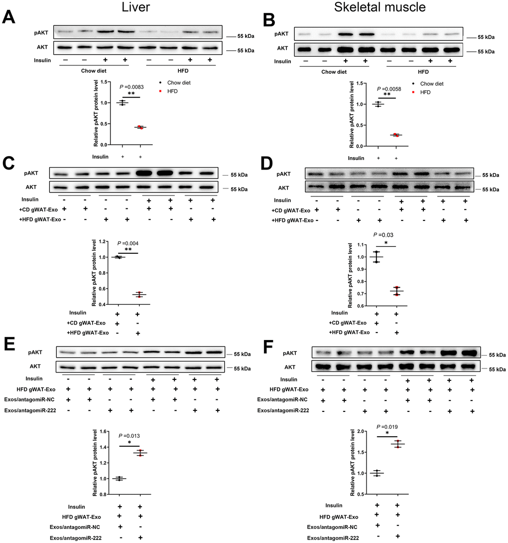
Figure 7.The gWAT-derived exosomal miR-222 impairs insulin signaling in the liver and skeletal muscle tissues of HFD-fed obese model mice. (A, B) Western blot analysis shows the phospho-AKT and AKT protein levels in the (A) liver and (B) skeletal muscle tissues from the CD-fed and HFD-fed mice. (C, D) Western blot analysis shows the phospho-AKT and AKT protein levels in the (C) liver and (D) skeletal muscle tissues in 8-week old wild-type mice continuously injected via the tail vein for 7 days with exosomes secreted by the adipose tissues from CD-fed or HFD-fed mice. (E, F) Western blot analysis shows the phospho-AKT and AKT levels in the (E) liver and (F) skeletal muscle tissues of mice continuously injected for 7 days via the tail vein with HFD-gWAT-derived exosomes plus 293T exosomes (containing antagomiR-NC or antagomiR-222). Note: For the in vivo insulin-stimulated AKT phosphorylation assay, the mice were injected with 0.75 IU/kg body weight insulin (i.p.) and sacrificed after 15 min. The phospho-AKT levels were normalized to the total AKT levels. The data are presented as the means ± SE; * P < 0.05, ** P < 0.01, *** P < 0.001.