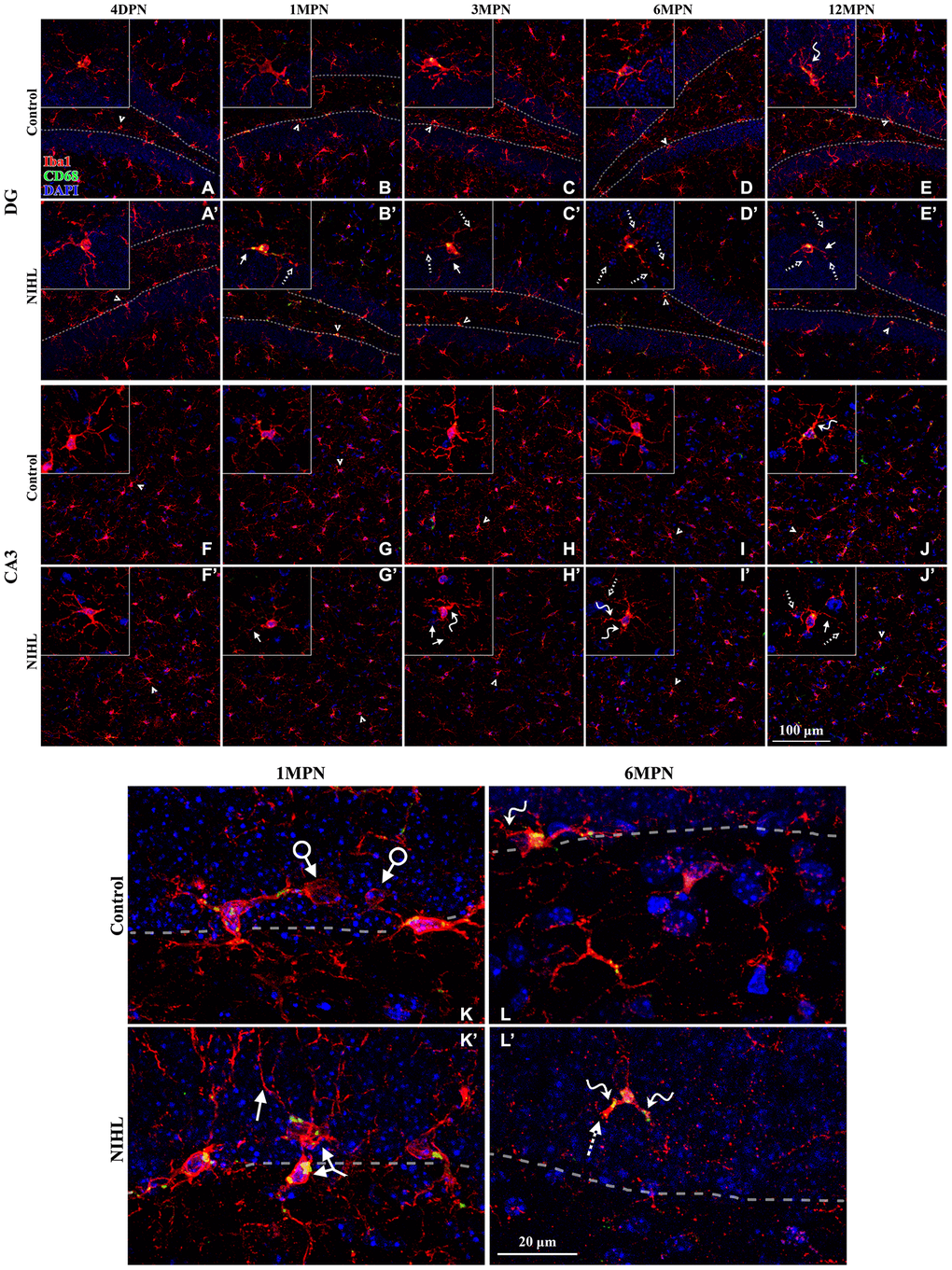
Figure 6.Microglial deterioration in the hippocampus associated with normal aging is aggravated by NIHL. (A–J’) Representative z-projection images of cells labeled with Iba1 (red), CD68 (green), and DAPI (blue) in the DG (A–E’) and CA3 (F–J’). Dotted lines in (A–E’) depict the SGZ (region between the granule cell layer and hilus). Iba1+ cells signified by arrowheads are magnified in the corresponding inserts. Scale bar equals 100 μm. (K–L’) Representative magnified images of Iba1-labeled cells in the DG from both groups at 1 MPN (K–K’) and 6 MPN (L–L’). Dotted lines depict the SGZ. Dystrophic cells are readily distinguished from ramified cells by degenerative changes in their cytoplasmic structure, such as deramified/atrophic cells (solid line arrows in B’, C’, E’, J’, H’, J’, and K’), fragmented or unusually tortuous processes (wavy arrows in E, J, H’, I’, L, and L’), and the formation of fragmented or beaded processes (dotted arrows in B’, C’, D’, E’, I’, J’, and L’). Arrows with circles in (K) point to microglial phagocytic pouches, a special modification of the microglial process involved in removing apoptotic cells from the adult SGZ neurogenic niche [27, 28]. The two-headed arrow in (K’) points to two abnormally aggregated microglial cells. Scale bar equals 20 μm.