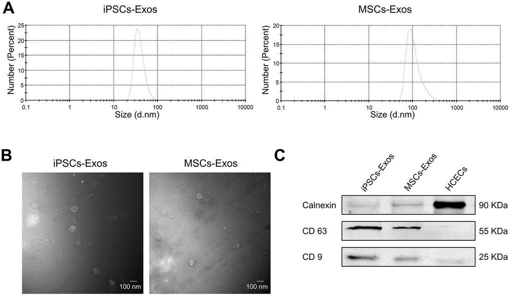
Figure 1.Characterization of iPSCs/MSCs-Exos. (A) NTA results demonstrate iPSCs/MSCs-Exos size distribution after isolation via ultracentrifugation. The diameter of iPSCs-Exos ranged from 30 to 120 nm while that of MSCs-Exos was between 60 and 400 nm. (B) TEM image of isolated exosomes showing a round morphology with diameters of approximately 100 nm. (C) Western blot illustrating the presence of the exosome markers CD9 and CD63, as well as the absence of the negative exosome markers Calnexin in isolated exosomes. HCEC cell lysate was used as a control.