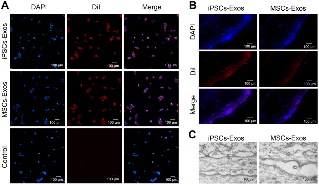
Figure 2.Uptake of iPSCs/MSCs-Exos. (A) Immunofluorescence staining of HCECs. HCECs were treated with 25 μg/ml DiI-labeled iPSCs/MSCs-Exos for 24 h. PBS, which was used to resuspend exosomes, was used as a negative control. Photomicrographs showed that red fluorescent particles were present throughout the cell cytoplasm, meaning that the exosomes were taken up by HCECs. (B) Immunofluorescence staining of rat corneal epithelium. After dropping DiI-labeled iPSCs/MSCs-Exos on rat cornea for 24 h, the corneas were harvested for immunofluorescence staining. The images of the whole mount of cornea showed a wide distribution of exosomes throughout the rat corneal epithelium, indicating successful fusion and uptake of iPSCs/MSCs-Exos by the corneal epithelium in vivo. (C) Exosome-like vesicles were detected by TEM on corneal epithelium. The diameters of the vesicles were between 100-200 nm.