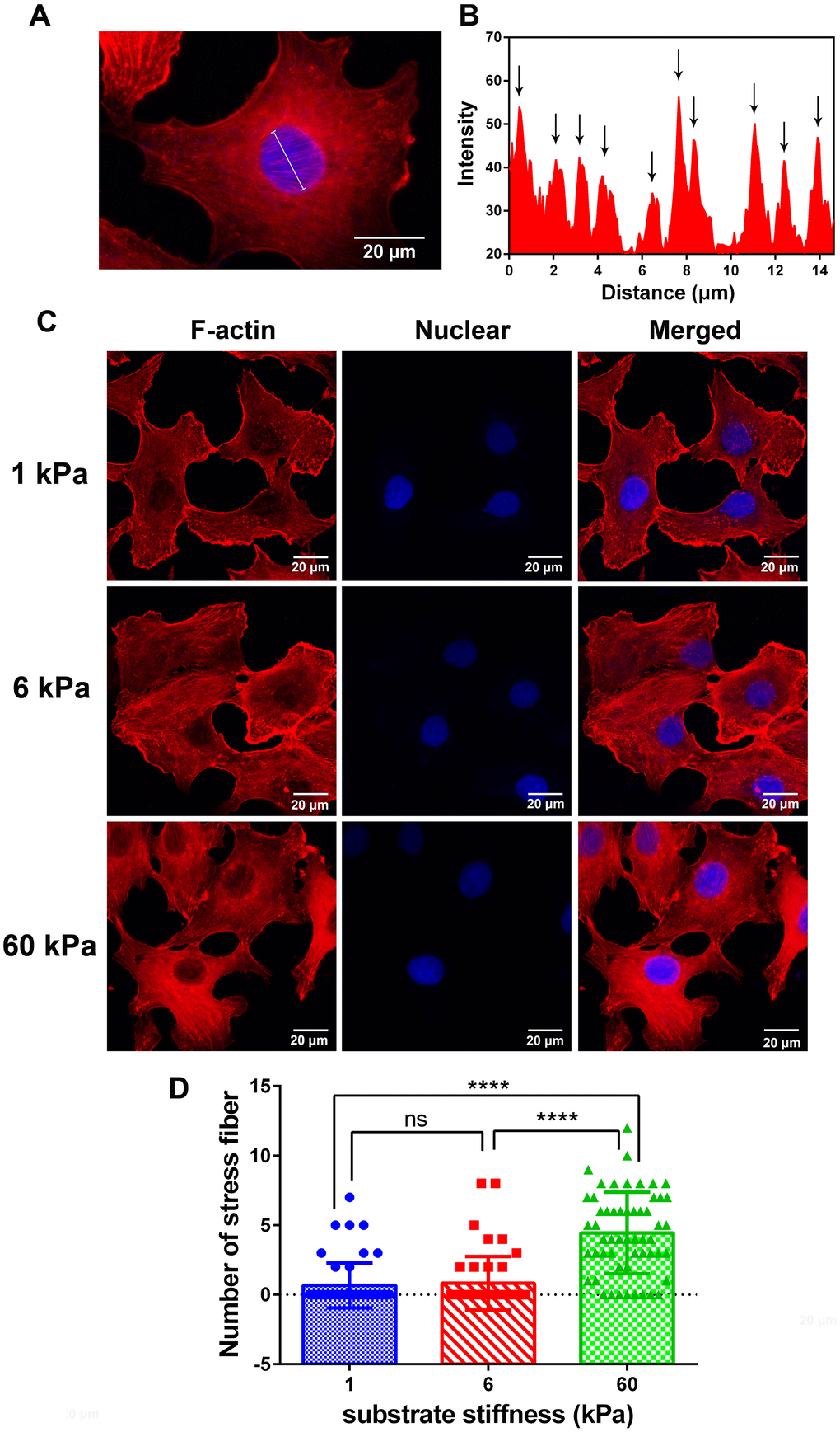
Figure 3.Immunofluorescence images and analysis of F-actin in SKOV-3 cells which grown on substrate with different rigidity. (A) Typical image was presented for defining the perinuclear actin stress fiber, and its analytic method using intensity calculated by ImageJ software (B). (C) SKOV-3 cells plated on collage I -coated 1 kPa, 6 kPa and 60 kPa hydrogels for 24 h were fixed and stained with rhodamine phalloidin (left) and DAPI (middle) to visualize F-actin and nucleus respectively. Scale bar - 20 μm. (D) Numbers of perinuclear actin stress fiber in SKOV-3 cells which grown on collage I-coated 1 kPa, 6 kPa and 60 kPa hydrogels for 24 h. Each column represents means ± SE of 49-55 cells from 2 experiments. **** P < 0.0001.