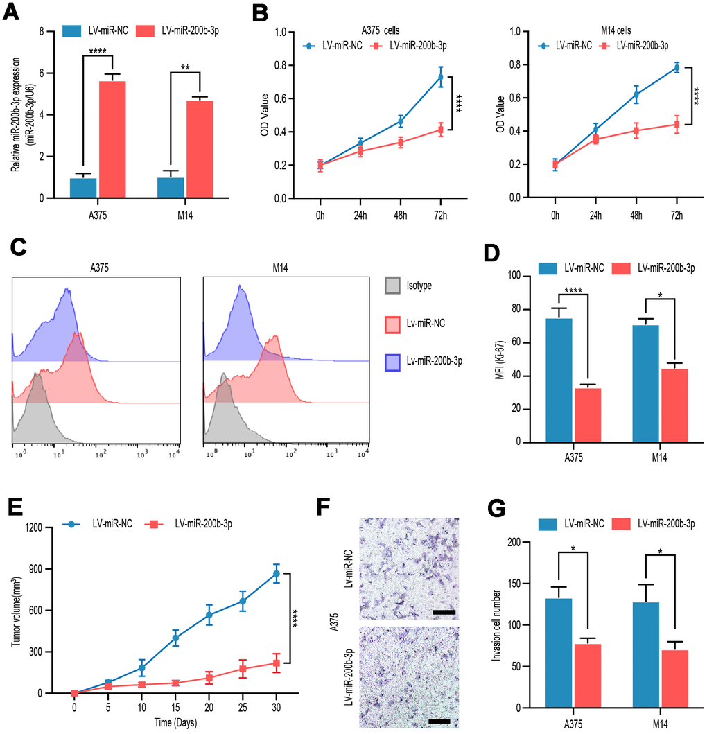
Figure 2.miR-200b-3p inhibited proliferation and invasion of melanoma cells. (A) A375 and M14 were transfected with LV-miR-NC or LV-miR-200b-3p for 24 hours and RT-qPCR was used to access miR-200b-3p levels. (B) CCK-8 assays were used to identify cell proliferation of LV-miR-200b-3p-transfected melanoma cells compared with that of control cells. (C–D) Following treatment for 48 hours, Ki67 was tested by flow cytometry in LV-miR-200b-3p-transfected cell lines compared with that of control cells. (E) Tumor growth curves were calculated after A375 cells transfected with miR-200b-3p. (F–G) Cell invasion was detected after cells transfected with LV-miR-200b-3p or control at 24h. Scale bars: 100 μm.