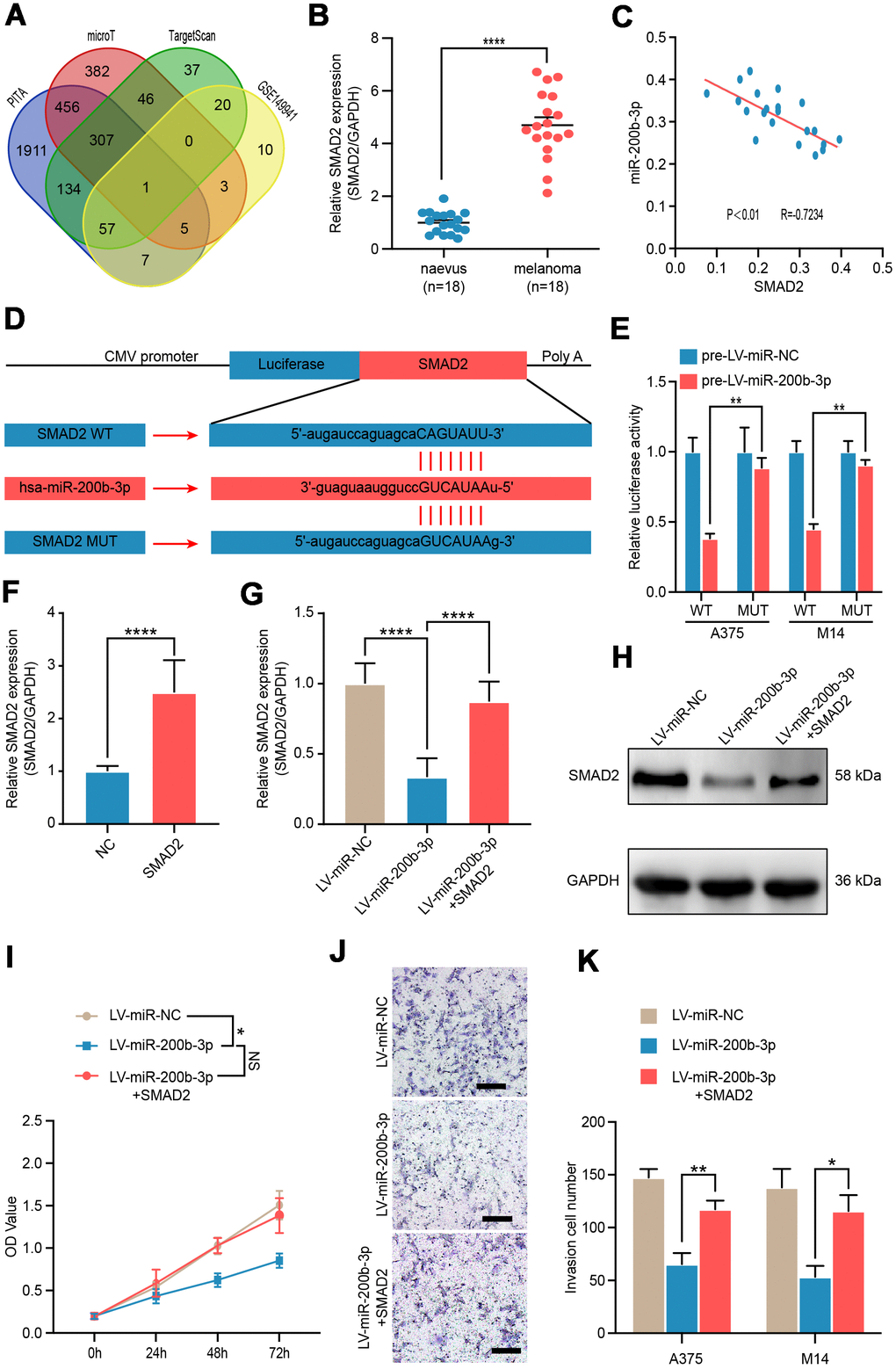
Figure 3.SMAD2 was verified as a functional target of miR-200b-3p. (A) Venn diagram of intersection of miRNA-200b-3p target genes predicted by bioinformatics analysis. (B) RT-qPCR was used to test expression of SMAD2 in benign nevus (n=18) and malignant melanoma tissues (n=18). (C) Spearman’s correlation analysis showed the correlation of miR-200b-3p and SMAD2 in malignant melanoma tissues (n=18). (D) Schematic view of putative miRNA-200b-3p targeting site in the Wt and Mut 3’-untranslated region (UTR) of SMAD2. (E) Luciferase reporter assay in A375 and M14 cells transfected with luciferase report plasmids containing SMAD2 3’- UTR (WT or MUT), and control miRNA or LV-miRNA-200b-3p. (F) RT-qPCR was used to test the efficiency of SMAD2 overexpression plasmid. (G–H) RT-qPCR and western blot were used to evaluate the mRNA and protein levels of SMAD2, after LV-miR-200b-3p and/or SMAD2 up-regulated lentivirus respectively. (I) CCK-8 assays were conducted to identify cell proliferation after cells were transfected LV-miR-NC, LV-miR-200b-3p, or LV-miR-200b-3p+SMAD2. (J–K) Cell invasion was detected after cells were transfected with LV-miR-NC, LV-miR-200b-3p, or LV-miR-200b-3p+SMAD2 or control at 24h. Scale bars: 100 μm.