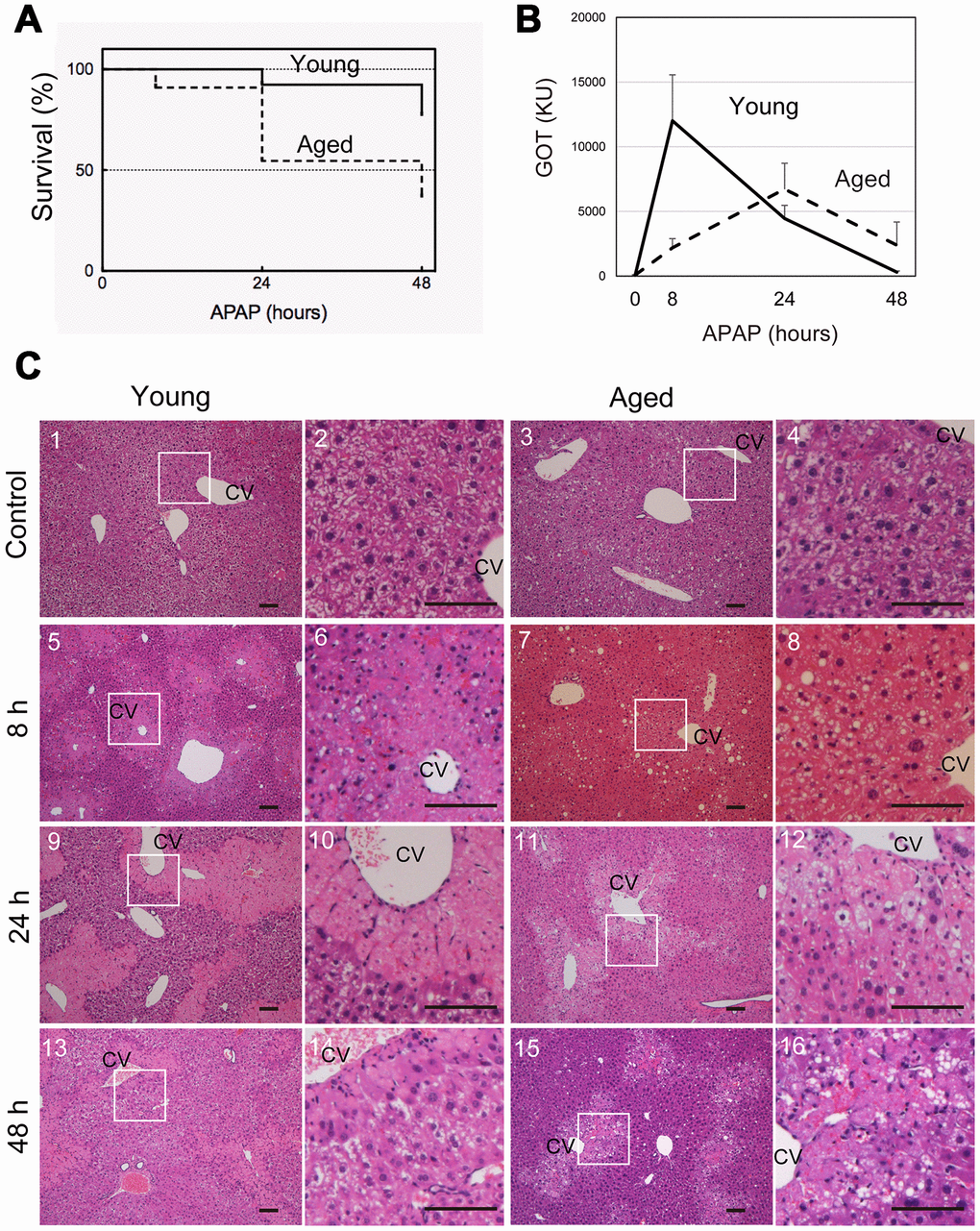
Figure 1.Aged mice are more susceptible to acetaminophen-induced liver injury. (A) Increased mortality by APAP injury in aged mice. Young (8-10W) and aged (>80W) mice were intraperitoneally injected with 300 mg/kg acetaminophen. N = 8 and 6 for young and aged mice, respectively. (B) Tissue damage is sustained at 48 hours after APAP injury in aged mice. Serum GOT increases at 8 hours after APAP administration, gradually decreases, and then gets back to the normal levels at 48 hours in young mice (solid line). By contrast, GOT is low at 8 hours and increases by 24 hours in aged mice (dotted line). GOT is still high at 48 hours. Serum was collected from more than six mice at each time point, and average values with SEMs are presented. (C) Histological analysis of young and aged liver tissue during APAP injury. APAP induces hepatocyte necrosis around CV, where hepatocytes lose their nuclei, and their cytoplasm is pale red with eosin staining (panels 5 and 6, 9 and 10). The damaged region is smaller at 48 hours after injury (panels 13 and 14). Contrastingly, in aged liver tissue, damaged areas are not clear at 8 hours (panels 7 and 8), become noticeable with pale eosin staining at 24 hours (panels 11 and 12), and persist at 48 hours (panels 15 and 16). Boxes in panels 1, 3, 5, 7, 9, 11, 13, and 15 are enlarged in panels 2, 4, 6, 8, 10, 12, 14, and 16, respectively. Bars represent 100 μm.