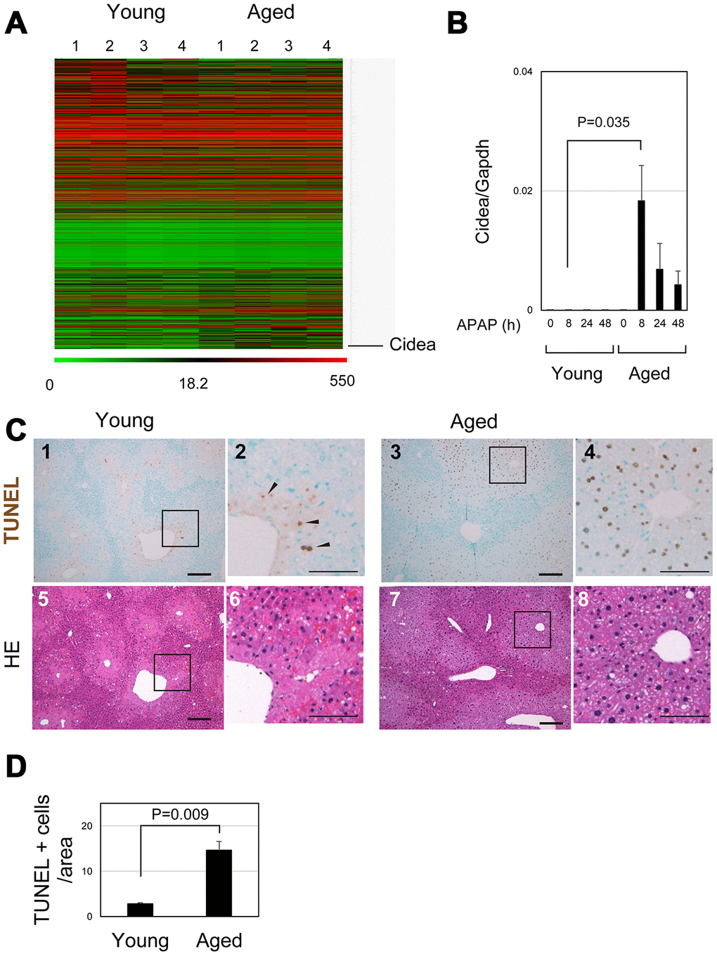
Figure 5.Apoptosis is promoted in aged liver. (A) Global gene expression profiles of young and aged livers at 8 hours after APAP injury. Cidea is listed as the most upregulated gene in aged livers 8 hours after APAP administration. Gene expression profiles were analyzed using microarray analysis. (B) Cidea is induced in aged liver tissue after APAP injury. Quantitative PCR analysis demonstrates that Cidea is significantly upregulated at 8 hours after APAP injury, specifically in aged livers. The graph shows average values with SEMs. (C) TUNEL staining of young and aged livers at 8 hours after APAP injury. APAP induces hepatocyte necrosis around the CV, and dead hepatocytes are mostly de-nucleated in young mice at 8 hours. In addition, a small number of TUNEL+ hepatocytes exist in the necrotic area. By contrast, hepatocytes around CV still possess their nuclei (panels 7 and 8), and they are mostly TUNEL+ (panels 3 and 4) in aged mice. Bars in panels 1, 3, 5, and 7, and panels 2, 4, 6, and 8 represent 50 and 100 μm, respectively. (D) Increase of TUNEL+ hepatocytes in aged livers. TUNEL+ hepatocytes within the distance of 100 μm from the CV is more in aged livers than those in young ones.