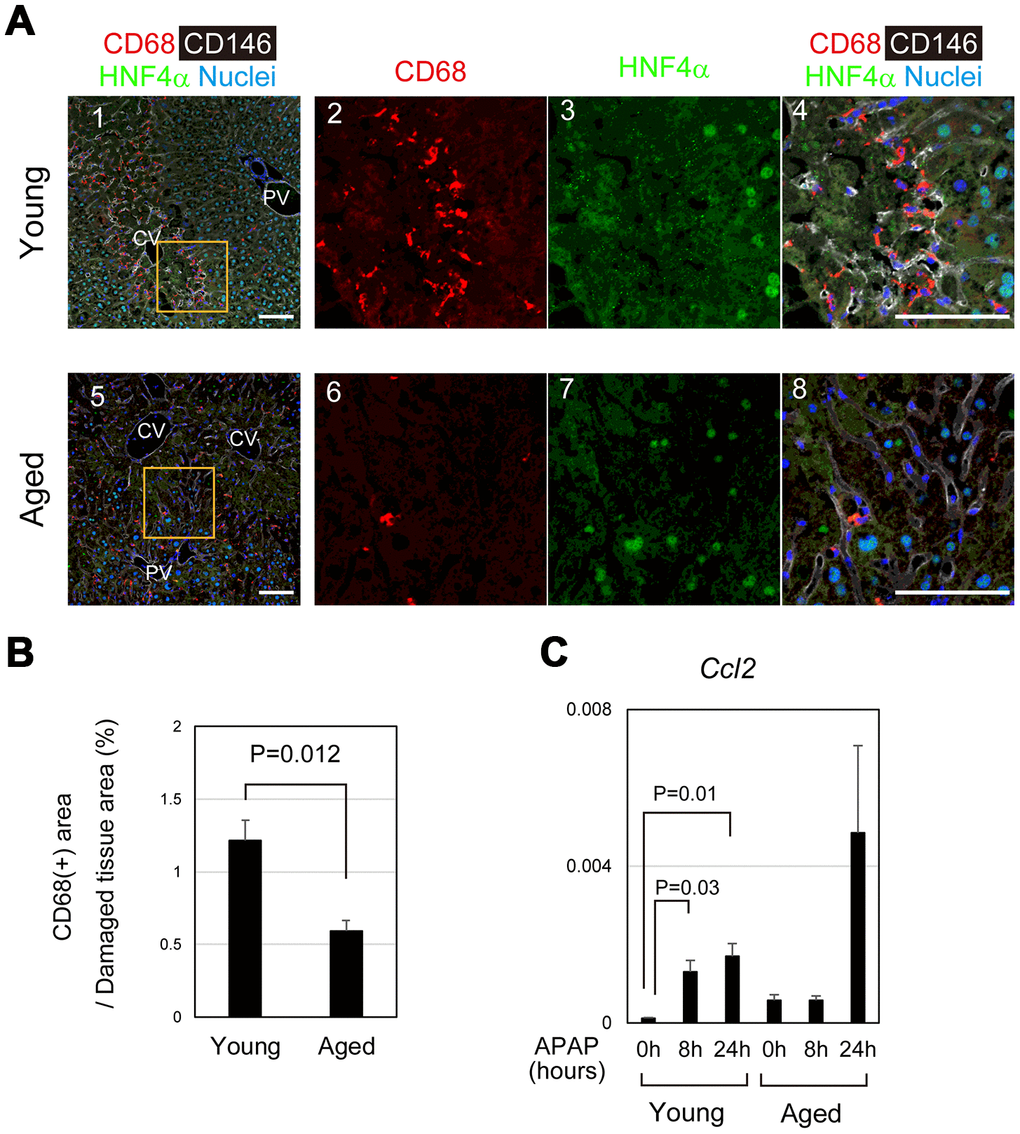
Figure 6.Impaired recruitment of macrophages in aged mice after APAP injury. (A) Macrophages accumulate toward the damaged area in young mice after APAP injury. CD68+ macrophages are abundant in damaged liver tissue around the CV at 24 hours in young mice (panels 1–4). Contrastingly, CD68+ macrophages in the damaged area of aged liver are minimal (panels 5–8). Scale bars in panels represent 100 μm. (B) Impaired recruitment of macrophages to damaged liver tissue in aged mice. CD68+ macrophages in the damaged tissue are significantly less in aged livers compared to those in young ones. Liver tissue consisting of HNF4α- hepatocytes was damaged by APAP administration. The recruitment of macrophages was estimated from the ratio of the CD68+ area in the HNF4α- tissue at 24 hours after APAP administration. Two areas were quantified on Image J. The graph shows average values with SEMs. (C) Induction of Ccl2 expression. Ccl2, chemokine-attracting macrophages, is upregulated in young mice at 8 and 24 hours after APAP administration with statistical significance. Conversely, it is not significantly induced in aged mice. The graph shows average values with SEMs.