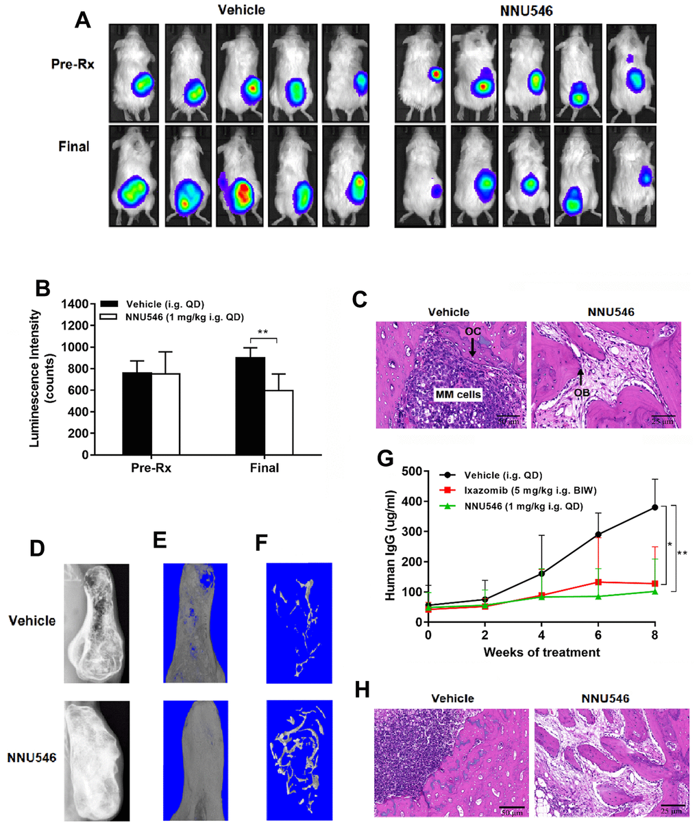
Figure 6.NNU546 inhibits the growth of human MM cells in SCID-rab mouse model. (A) Rabbit bone grafts were subcutaneously implanted into SCID mice. After four weeks, 5×10⁶ of RPMI 8226 cells with luciferase expression were injected directly into the rabbit bone in SCID mice. After establishment of this in vivo model, mice were orally administered with either vehicle or NNU546 (1 mg/kg, QD). Representative imaging of the live animals demonstrated the luciferase expression prior to treatment (Pre-Rx) and at the end of the experiment (Final) in mice. (B) Luminescence intensity was quantified using living animal imaging of mice prior to treatment (Pre-Rx) and at the end of the experiment (Final). Data were expressed as mean ± SD (n=5; **, p < 0.01). (C) H&E staining of histological sections of myelomatous bones engrafted with myeloma cells (×200 or ×400 original magnification). Increased myeloma cell infiltration and osteoclast activity were noted in control group. In contrast, myelomatous bone from a host treated with NNU546 had no apparent myeloma cells but possessed increased number of trabecular bone and osteoblast. (D) SCID-rab mice engrafted with myeloma cells from MM patients were treated with vehicle, ixazomib (5 mg/kg, BIW) or NNU546 (1 mg/kg, QD). X-radiographs of myelomatous bones engrafted with myeloma cells from MM patients were performed at the end of the experiment. (E, F) Representative microCT images of bone grafts excised from vehicle-treated animals and mice that had received NNU546 for 8 weeks. (G) Effect of therapy on myeloma growth assessed by human IgG levels in the mouse serum measured by ELISA. Data were shown as mean ± SD (n=5; *, p < 0.05; **, p < 0.01). (H) H&E staining of histological sections of myelomatous bones engrafted with cells from MM patients (original magnification, ×200 or ×400 original magnification).