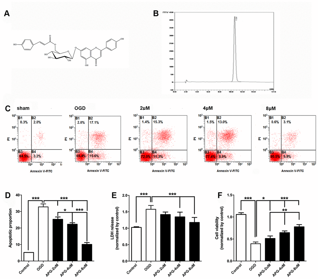
Figure 1.Protective effect of APG against OGD/R injury in cortical neurons. (A) Chemical structure of APG (molecular weight: 578; molecular formula: C30H26O12). (B) HPLC analysis of APG (retention time = 11.047 min). The detection wavelength was 254 nm; the mobile phase was methanol and 0.1% phosphoric acid at a ratio of 40:60 (v/v) through the elution. The flow rate was kept at 1 mL/min. (C) Representative dot plots showing flow cytometric analysis of cortical neurons treated with different concentrations of APG and then stained with FITC-conjugated Annexin V and propidium iodide in control, OGD, 2 μM, 4 μM and 8 μM APG treatment groups. (D) Analysis result of apoptotic index in each group (n = 5). (E) Effect of APG treatment on plasma lactate dehydrogenase (LDH) releasing level in primary cortical neuron culture challenged by OGD/R. (F) Effect of APG treatment on cell viability in primary cortical neuron culture subjected to OGD/R. n = 5 per group. * P < 0.05, ** P < 0.01, *** P < 0.001, respectively.